
Page 63 - Read Online
P. 63
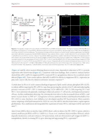
Page 8 of 19                                                   Lin et al. Cancer Drug Resist. 2026;9:14


               Figure 3. Therapeutic concept and in vivo effects of mPTEN@NPs in a B16F10 melanoma model. This figure illustrates mPTEN@NPs as a
               nanoplatform designed to restore PTEN-related antitumor activity and remodel the immunosuppressive TME, thereby helping to
               overcome resistance to immunotherapy. The proposed strategy enhances immune activation while reducing suppressive mechanisms
               associated with tumor progression. Model: B16F10 melanoma. Key readouts: tumor control, lymph node dendritic cell activation, CD8 +
               T-cell infiltration and effector function, reduced Treg and Mo-MDSC populations, cytokine modulation, PTEN and LC3-II expression, and
               ATP release. (A) Experimental timeline; (B) Tumor weights of B16F10 tumor-bearing mice treated with PTEN@NPs; (C and D) Flow
               cytometric analysis of the percentages of Foxp3  CD25  CD4  T cells (C) and Mo-MDSCs (D); (E) Immunofluorescence imaging of PTEN
                                               +
                                                       +
                                                   +
               (green) and LC3-II (red) in PTEN-mutated B16F10 tumor tissues. Adapted with permission from American Association for the
               Advancement of Science [62] . mPTEN: PTEN mRNA (PTEN: phosphatase and tensin homolog); NPs: nanoparticles; TME: tumor
               microenvironment; Treg: regulatory T cell; Mo-MDSC: monocytic myeloid-derived suppressor cell; ATP: adenosine triphosphate.
               [Figure 4A and B], while western blotting demonstrated a time-dependent reduction in PD-L1 protein
               expression after transfection [Figure 4C]. Consistent with these findings, flow cytometric analysis further
               showed that siPD-L1@PLGA suppressed IFN-γ-induced PD-L1 upregulation, whereas the scrambled control
               did not [Figure 4D]. Their results indicate that siPD-L1@PLGA effectively suppresses PD-L1 expression in
               pancreatic cancer cells, thereby enhances antitumor immune responses .
                                                                          [64]
               A study done by Wu et al. used a nanotechnological approach, lipid-coated calcium phosphate NPs (LCN),
               to deliver siRNA targeting the PD-1/PD-L1 axis, thus preserving the activity of the T cells and reducing the
               primary resistance of PD-1/PD-L1 immunotherapy. LCN-siRNA PD-1/PD-L1 NPs targeting PD-1 and
               PD-L1 were administered separately, resulting in enhanced cytotoxic T-cell activity, and improved antitumor
               efficacy. In this combination silencing, the cytotoxicity was related to the increased release of IFN-γ and
               tumor necrosis factor-α (TNF-α) . Upregulation of these cytokines further contributed to blockade of the
                                           [65]
               PD-1/PD-L1 axis. Similarly, Erel-Akbaba et al. developed another NP-based system. They created a
               tumor-targeting solid lipid nanoparticle (SLN) to carry the siRNA into the brain tumor region against
               glioblastoma. This combination downregulated the expression of tumor PD-L1 and improved the survival of
               mice .
                   [66]

               Apart from siRNA, there is another type of RNA that is able to silence the PD-1/PD-L1 genes, which is
               named small hairpin RNA of PD-L1 (shPD-L1). Guan et al. combined hyaluronidase (HAase), which
               degrades hyaluronic acid and enhances NP penetration, with an ultrasensitive pH-triggered shPD-L1
               nanoplatform, thereby increasing PD-L1 gene silencing and suppressing PD-L1 expression .
                                                                                           [67]


                                                           56