
Page 128 - Read Online
P. 128
Page 8 of 13 Fang et al. Cancer Drug Resist. 2025;8:42
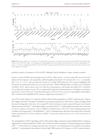
Figure 4. Molecular differences in immune cell composition and immune-related gene expression between ROS1-WT and ROS1-Mut
tumors in the TCGA HNC cohort. (A) Comparison of 22 predefined immune cell types; (B) expression of 39 immune-related genes. P <
*
0.05; P < 0.01; ns: not significant (P ≥ 0.05). ROS1-WT: ROS1-wild-type; ROS1-Mut: ROS1 mutations; TCGA: The Cancer Genome Atlas;
**
HNC: head and neck cancer.
predictive marker of resistance to ICIs in HNC, although clinical validation in larger cohorts is needed.
Despite elevated TMB and neoantigen load in ROS1-Mut tumors - features typically associated with
enhanced ICI response - these patients exhibited significantly shorter OS (median OS: 5.0 vs. 11.0 months,
HR = 3.22, P = 0.011). The paradoxical coexistence of high TMB with poor ICI response highlights the
limitations of mutational burden as a universal biomarker. While TMB generally correlates with neoantigen
visibility, ROS1-Mut tumors may override this immunogenic advantage through MYC-mediated
transcriptional reprogramming. This is supported by significant downregulation of antigen presentation
machinery (TAP1/TAP2) and T cell chemotaxis genes (CXCL9/CXCL10) in our transcriptomic data [Figure
4B], consistent with established MYC-immune suppression mechanism [27-32] .
Our GSEA results [Figure 5A], together with the observed downregulation of MHC-I pathway genes [Figure
4B], suggest that MYC activation contributes to an immunosuppressive TME where a high neoantigen load
becomes biologically irrelevant, mirroring MYC-driven immune evasion reported in pancreatic ductal
adenocarcinoma (PDAC) and hepatocellular carcinoma (HCC) . Similar phenomena have been observed
[32]
[31]
in other oncogenic alterations, such as MDM2/4 amplification, where ICI-induced hyperprogression
correlates with suppressed T cell infiltration . JAK1/2 mutations have also been associated with accelerated
[27]
tumor growth post-ICI due to defective MHC-I expression and resistance to T cell cytotoxicity in
melanoma .
[28]
The upregulation of MYC signaling in ROS1-Mut tumors aligns with prior studies linking MYC to immune
suppression via downregulation of MHC class I molecules and recruitment of immunosuppressive myeloid
cells [29-32] . These defects in antigen presentation represent a rate-limiting step that impedes neoantigen
visibility , explaining the discordance between high TMB and poor ICI response in ROS1-mutant tumors.
[33]
121