
Page 47 - Read Online
P. 47
Villavisanis et al. Art Int Surg. 2025;5:133-38 https://dx.doi.org/10.20517/ais.2024.89 Page 135
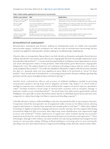
Table 1. Salient studies applying AI to microvascular reconstruction
Author, year, journal Aim Implications
Asaad et al., 2023, Annals Determine factors predictive of head and neck Machine learning models determined predictors of flap
[22]
of Surgical Oncology microvascular flap failure complications, most commonly smoking, flap type, and vein graft
Kuo et al., 2018, Use neural networks to predict surgical site Neural networks were more predictive than logistic regression for
[35]
Oncotarget infection after head and neck free flap surgical site infections after head and neck microvascular
reconstruction reconstruction
Kim et al., 2024, Develop an AI-based automated free flap An AI-based flap monitoring system may reduce postoperative
JAMA Network [33] monitoring system via evaluation of clinical clinician burden and workload
photography
O’Neill et al., 2020, Develop a machine learning model that can Machine learning model identified high-risk patient factors
Annals of Surgical predict flap failure from a large clinical dataset including obesity, comorbidities, and smoking
Oncology [34]
INTRAOPERATIVE ASSESSMENT
Intraoperative assessment and decision making are fundamental parts of reliable and repeatable
microvascular surgery. Artificial intelligence not only has roles in intraoperative monitoring, but has
recently been applied to intraoperative decision making for troubleshooting complications.
Objective data on intraoperative flap perfusion can help identify perforasomes and guide clinical decision
making. Specifically, indocyanine green fluorescence angiography has been increasingly employed to assess
[25]
intraoperative flap perfusion . A recent study leveraged artificial intelligence-based applications to review
and assess intraoperative videos of flap perfusion with indocyanine green fluorescence angiography
(Singaravelu 2024). The authors found over 99% validation and testing accuracy with the need to retain or
excise peripheral flap portions . The study also identified a threshold of regions with fluorescence intensity
[26]
less than 22.1 grayscale units that were significantly more likely to be predicted as “excise” by these
models . Such models may be beneficial in corroborating intraoperative decision making regarding flap
[26]
perfusion and the extent of peripheral tissue included in the flap .
[26]
Another study evaluated the efficacy and accuracy of artificial intelligence models in providing
intraoperative guidance during deep inferior epigastric perforator flap surgery , in which artificial
[27]
intelligence responses were evaluated by board-certified plastic surgeons on several objective, quantitative
scales . Prompts included a broad range of intraoperative scenarios such as iatrogenic damage to
[27]
perforator vessels or acute arterial thrombosis . The study found that while answers generated by artificial
[27]
intelligence were generally accurate, they lacked nuance specific to individual patient factors and were more
comparable to resident knowledge level than to experienced attending surgeons .
[27]
Critically relevant to trainees, artificial intelligence has also demonstrated utility in microsurgery education.
Groups have trialed the intraoperative use of augmented reality overlays in free fibula harvest, allowing
surgeons and trainees to visualize bony anatomy and vascular paths in real time [28,29] . Others have utilized
technology to digitize preoperative imaging and project vascular anatomy into the surgical field during
anterolateral thigh free flap procedures, thus improving vessel identification with impressively high
accuracy and sensitivity . Similar applications of augmented reality headsets during microvascular
[30]
anastomosis performed by resident trainees demonstrated improved visualization and ergonomics . These
[31]
technologies, although in their infancy, can assist residents not only during simulated microsurgery but also
during the intraoperative setting. Utilizing artificial intelligence technology prior to even entering the
operating room may yield improvements to surgical understanding, trainee dexterity, and procedure safety.