
Page 67 - Read Online
P. 67
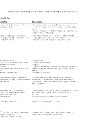
Page 160                                                                                                                                                      Kimbowa et al. Art Int Surg 2024;4:149-69  https://dx.doi.org/10.20517/ais.2024.20


 Table 2. A summary of the deep reviewed learning-based methods listed in descending order of date published

 Date  Author                     Mode Approach  Strengths  Limitations
 [87]
 2024-  Che et al.  2D  - Calibrate ultrasound probe and needle to optical tracker   - Combines both hardware and software-  - Requires extra hardware: optical tracker and localizers
 01-12  - Capture needle tip using tracker   based approaches  - Calibration may be erratic if the tip strays from the imaging
 - Represent tip in the ultrasound image coordinate system  plane
                                        - Involves a two-person workflow and separate algorithms for
                                        needle tracking and detection
 [91]
 2023-  Yan et al.  2D+t  Consists of 3 modules   - Incorporates temporal information   - Fails if there is sustained disappearance of the needle tip
 05-17  (1) Visual tracking module   - Thoroughly evaluated for multiple users   - Tracking fails when the needle tip is too small and
 - Pass sampled patches from consecutive frames through the 2- and insertion motions  indistinguishable from background right at insertion
 branch Siamese network
 - One branch dynamically extracts features from template
 patches and the other extracts features from current patch
 (2) Motion prediction module
 - Needle velocity is estimated using displacement of the tip
 between consecutive frames
 (3) Data fusion
 - Use MLP to independently fuse data from visual tracking
 module and motion prediction module
 [84]
 2023-  Wang et al.  2D+t  (1) Enhanced ultrasound image by subtracting current frame   - Accounts for motion   - Only in plane
 05-09  from previous frame and fusing the difference with the current   - Continous needle detection even when   - Detects only needle tip
 frame   static between frames          - In plane
 (2) Extract spatial constraint (bounding box around shaft) if   - Real time   - Mentions that different detectors could be used, but this is
 present   - Evaluated on human data- Evaluated on   not true as specific detectors (that yield bounding boxes)
 (3) Extract temporal constraint (bounding box around tip) if   CPU   should be used
 present   - Independent of detector    - Doesn’t visualize shaft
 (4) Combine the constraints to localize needle tip
 [88]
 2023-  Zade et al.  2D+t  - Extract motion field’s amplitude and phase from successive   - Evaluated approach on 2 categories of   - No ablation study to show relevance of the different
 02-06  frames   images (needle aligned correctly, and needle  components of the proposed assisted excitation module
 - Extract spatiotemporal features from the motion fields   imperceptible)  - No comparison with SOTA to show how they fail to model
 - Pass features through detector that outputs a vector of size   speckle dynamics
 values including pixelwise line parameters (tx,ty,theta), and
 probabilities for needle shaft and tip
 [78]
 2021-  Chen et al.  2D  - 2 frameworks   - Segments shaft, localizes needle   - Does not justify how approach accounts for time (inputs are
 10-22  (1) Predict segmentation from two adjacent images   - Doesn’t require prior knowledge of   two consecutive images but with no time information
 (2) 2 models - one segments shaft, the other segments needle   insertion side/orientation   encoded)
 tip from ROIs extracted from the predictions of (1)   - Fully automatic  - Did not compare with state of the art methods
 - Use segmentations to find
 [77]
 2021-  Wijata et al.  2D  - Pass image through Unet like architecture   - Evaluated on in vivo data  - Not robust to high intensity artifacts
 06-28  - Architecture uses large kernels (11 × 11, 9 × 9, 7 × 7) to detect
 large objects
 - Apply Radon transform to determine needle trajectory
 - back-transform trajectory back to binary image
 [85]
 2021-  Rubin et al.  3D  - Pass k previous frames to 3D CNN (including current frame)   - Efficient (real-time, can run on low-cost   - Only detects needle with a bounding box and does not
 05-11  - Repeatedly apply 3D convolution to obtain a single feature   computing hardware)   provide metrics on needle tip localization
 map for temporal information   - Evaluated on challenging cases
 - Pass the feature map through a 2D detector
 - 2D detector outputs bounding box of needle in current frame