
Page 62 - Read Online
P. 62
Page 156 Kimbowa et al. Art Int Surg 2024;4:149-69 https://dx.doi.org/10.20517/ais.2024.20
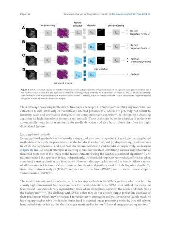
Figure 3. Software-based needle localization methods can be categorized into three: (A) classical image processing methods that use a
handcrafted feature extractor and decoder; (B) machine learning-based methods with a trainable decoder; (C and D) and deep learning-
based methods with a trainable feature extractor and decoder. Generally, software-based methods take as input either single ultrasound
images or a video stream of ultrasound images.
Classical image processing methods face two major challenges: (1) they require carefully engineered feature
extractors E with arbitrarily or heuristically selected parameters λ which are generally not robust to
1
[63]
intensity, scale and orientation changes, or are computationally expensive ; (2) designing a decoding
algorithm for high dimensional features is not tractable. These challenges led to the adoption of methods to
automatically learn features necessary for needle detection and also learn robust classifiers for high-
dimensional features.
Learning-based methods
Learning-based methods can be broadly categorized into two categories: (1) machine learning-based
methods in which only the parameters λ of the decoder D are learned, and (2) deep learning-based methods
2
in which the parameters λ and λ of both the feature extractor E and decoder D, respectively, are learned
1
2
[Figure 3B and C]. Initial attempts at learning a classifier involved combining various combinations of
threshold responses of the image to the feature extractors using the Adaboost statistical algorithm . The
[67]
intuition behind this approach is that, independently, the threshold responses are weak classifiers, but when
combined, a strong classifier can be obtained. However, this approach is wasteful as it only utilizes a subset
of all the extracted features. Other common classification algorithms used include Bayesian classifer ,
[68]
[56]
linear discriminant analysis (LDA) , support vector machine (SVM) , and its variant linear support
[60]
vector machine (LSVM) .
[56]
The most commonly used decoder in machine learning methods is the SVM algorithm, which can learn to
classify high-dimensional features from data. For needle detection, the SVM is fed with all the extracted
features and it outputs a binary segmentation mask where white pixels represent the needle and black pixels
the background [55,60,69] . The challenge with SVMs is that they do not directly output probability estimates of
their predictions which may be required for uncertainty estimation and postprocessing. While machine
learning approaches solve the decoder issues faced in classical image processing methods, they still rely on
handcrafted features that exhibit the challenges mentioned in Section “Classical image processing methods”.