
Page 65 - Read Online
P. 65
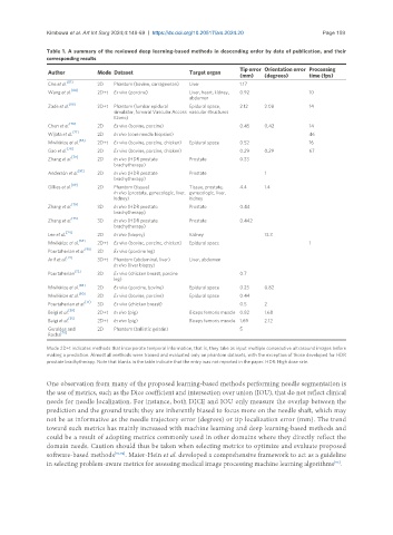
Kimbowa et al. Art Int Surg 2024;4:149-69  https://dx.doi.org/10.20517/ais.2024.20                                                      Page 159

               Table 1. A summary of the reviewed deep learning-based methods in descending order by date of publication, and their
               corresponding results
                                                                          Tip error Orientation error  Processing
                Author                          Mode Dataset  Target organ
                                                                          (mm)   (degrees)     time (fps)
                     [87]
                Che et al.     2D   Phantom (bovine, carrageenan)  Liver  1.17
                Wang et al. [84]  2D+t  Ex vivo (porcine)  Liver, heart, kidney,   0.92        10
                                                          abdomen
                      [88]
                Zade et al.    2D+t  Phantom (lumbar epidural   Epidural space,   2.12  2.08   14
                                    simulator, femoral Vascular Access  vascular structures
                                    Ezono)
                Chen et al. [78]  2D  Ex vivo (bovine, porcine)           0.45   0.42          14
                       [77]
                Wijata et al.  2D   In vivo (core needle biopsies)                             46
                Mwikirize et al. [66]  2D+t  Ex vivo (bovine, porcine, chicken)  Epidural space  0.52  16
                     [76]
                Gao et al.     2D   Ex vivo (bovine, porcine, chicken)    0.29   0.29          67
                Zhang et al. [79]  2D  In vivo (HDR prostate   Prostate   0.33
                                    brachytherapy)
                Andersén et al. [81]  2D  In vivo (HDR prostate   Prostate       1
                                    brachytherapy)
                       [89]
                Gillies et al.  2D  Phantom (tissue)      Tissue, prostate,   4.4  1.4
                                    In vivo (prostate, gynecologic, liver,  gynecologic, liver,
                                    kidney)               kidney
                Zhang et al. [79]  3D  In vivo (HDR prostate   Prostate   0.44
                                    brachytherapy)
                Zhang et al. [75]  3D  In vivo (HDR prostate   Prostate   0.442
                                    brachytherapy)
                Lee et al. [74]  2D  In vivo (biopsy)     Kidney                 13.3
                         [65]
                Mwikirize et al.  2D+t  Ex vivo (bovine, porcine, chicken)  Epidural space     1
                Pourtaherian et al. [90]  3D  Ex vivo (porcine leg)
                     [73]
                Arif et al.    3D+t  Phantom (abdominal, liver)   Liver, abdomen
                                    In vivo (liver biopsy)
                        [72]
                Pourtaherian   3D   Ex vivo (chicken breast, porcine      0.7
                                    leg)
                Mwikirize et al. [83]  2D  Ex vivo (porcine, bovine)  Epidural space  0.23  0.82
                         [60]
                Mwikirize et al.  3D  Ex vivo (bovine, porcine)  Epidural space  0.44
                Pourtaherian et al. [71]  3D  Ex vivo (chicken breast)    0.5    2
                      [69]
                Beigi et al.   2D+t  In vivo (pig)        Biceps femoris muscle 0.82  1.68
                Beigi et al. [55]  2D+t  In vivo (pig)    Biceps femoris muscle 1.69  2.12
                Geraldes and   2D   Phantom (ballistic gelatin)           5
                Rocha [70]
               Mode 2D+t indicates methods that incorporate temporal information, that is, they take as input multiple consecutive ultrasound images before
               making a prediction. Almost all methods were trained and evaluated only on phantom datasets, with the exception of those developed for HDR
               prostate brachytherapy. Note that blanks in the table indicate that the entry was not reported in the paper. HDR: High dose rate.


               One observation from many of the proposed learning-based methods performing needle segmentation is
               the use of metrics, such as the Dice coefficient and intersection over union (IOU), that do not reflect clinical
               needs for needle localization. For instance, both DICE and IOU only measure the overlap between the
               prediction and the ground truth; they are inherently biased to focus more on the needle shaft, which may
               not be as informative as the needle trajectory error (degrees) or tip localization error (mm). The trend
               toward such metrics has mainly increased with machine learning and deep learning-based methods and
               could be a result of adopting metrics commonly used in other domains where they directly reflect the
               domain needs. Caution should thus be taken when selecting metrics to optimize and evaluate proposed
               software-based methods [93,94] . Maier-Hein et al. developed a comprehensive framework to act as a guideline
                                                                                                      [93]
               in selecting problem-aware metrics for assessing medical image processing machine learning algorithms .