
Page 59 - Read Online
P. 59
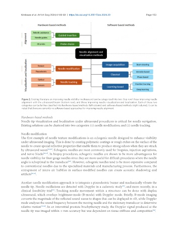
Kimbowa et al. Art Int Surg 2024;4:149-69  https://dx.doi.org/10.20517/ais.2024.20                                                      Page 153


                Figure 2. Existing literature on improving needle visibility in ultrasound can be categorized into two: (top row) those improving needle
                alignment with the ultrasound beam (bottom row), and those improving needle visualization and localization. Each of these two
                categories can be further classified into hardware-based methods (left column) and software-based methods (right column). It can be
                noted that there are currently no software-based approaches for improving needle alignment.

               Hardware-based methods
               Needle tip visualization and localization under ultrasound procedures is critical for needle navigation.
               Existing solutions can be clustered into two categories: (1) needle modification; and (2) needle tracking.

               Needle modification
               The first example of needle texture modifications is an echogenic needle designed to enhance visibility
               under ultrasound imaging. This is done by creating polymeric coatings or rough etches on the surface of the
               needle to create special reflective properties that enable them to produce strong echoes when they are struck
               by ultrasound waves [29,30] . Echogenic needles are most commonly used for biopsies, injection aspirations,
               and nerve blocks [30,31] . In biopsy procedures, echogenic needles are shown to be more advantageous for
               needle visibility for finer gauge needles since they are more useful for difficult procedures where the needle
               angle is suboptimal to the transducer . However, echogenic needles tend to be more expensive compared
                                               [29]
               to conventional needles due to the specialized materials and manufacturing process. Furthermore, the
               entrapment of micro air bubbles in surface-modified needles can create acoustic shadowing and
               artifacts [29,32] .

               Another needle modification approach is to integrate a piezoelectric buzzer and mechanically vibrate the
               needle tip. Needle oscillations are detected with Doppler in a cadaveric study , and more recently, in a
                                                                                   [33]
               clinical feasibility trial . Tracking needle movement within a structure can be done with duplex
                                    [34]
               ultrasound, which overlays brightness-mode (B-mode) with Doppler mode. Briefly, B-mode imaging
               converts the magnitude of the reflected sound waves to shapes that can be displayed in 2D, while Doppler
               mode analyzes the sound frequency between the moving needle and the stationary transducer to determine
               relative motion [35,36] . In an interstitial prostate brachytherapy study, the Doppler signal produced at the
               needle tip was imaged within 1-mm accuracy but was dependent on tissue stiffness and composition .
                                                                                                       [34]