
Page 112 - Read Online
P. 112
Jiao et al. Art Int Surg 2023;3:98-110 https://dx.doi.org/10.20517/ais.2023.03 Page 104
bringing the jejunum over to the right side after Kocherization of the duodenum. The mesentery to the first
15 cm of jejunum is divided close to the mesenteric border using the vessel sealer in arm 3. Then, the
jejunum is marked 80 cm from the DJ flexure with 2/0 PDS and brought over to the supracolic
compartment and anchored to the anterior abdominal wall in front of the stomach. This step effectively
reduces the back-and-forth movement of instruments during later stages of the operation, reducing
operative time. Now the camera along with instruments are moved towards the right side of the mesentery
and the jejunum is pulled through the retro mesenteric window followed by jejunal transection around 15
cm distal to DJ flexure using the stapler (Echelon Flex Powered plus articulating endoscopic linear cutter 60
mm, Ethicon, USA).
Pancreatic tunneling and resection of the pancreatic neck
During this step, the inferior border of the neck of the pancreas is dissected off the mesentery with hook
diathermy to identify and expose the SMV. Subsequently, gentle dissection is carried out behind the
pancreatic neck using hook diathermy in combination with mild irrigation and suction by the assistant.
Care is taken to avoid bleeding from the venous tributaries to the portal vein during the tunneling behind
the pancreatic neck. Dissection at the angle between the CHA and GDA will expose the origin of the PV
above the superior border of the pancreatic head. After completion of pancreatic tunneling above the SMV,
the neck of the pancreas is divided using the vessel sealer, and hemostasis of the pancreatic remnant is
achieved with the use of bipolar fenestrated grasper in arm 1. In the case of inflammatory adhesions around
the pancreas, dissection proceeds cranially from the route of the mesentery along the SMV to complete
tunneling of the pancreas.
Transection of the common hepatic duct and cholecystectomy
Following pancreatic neck transection, the common bile duct is dissected from the proper hepatic artery
using hook diathermy after the application of Hem-o-loks to small blood vessels. Then, the gallbladder is
mobilized from the liver bed and the common hepatic duct (CHD) is dissected from the hepatic artery and
PV followed by transection of the CHD with hook diathermy. Subsequently, the bile duct along with retro
portal and superior pancreaticoduodenal nodes are mobilized from the portal vein to the level of the origin
of the SMA.
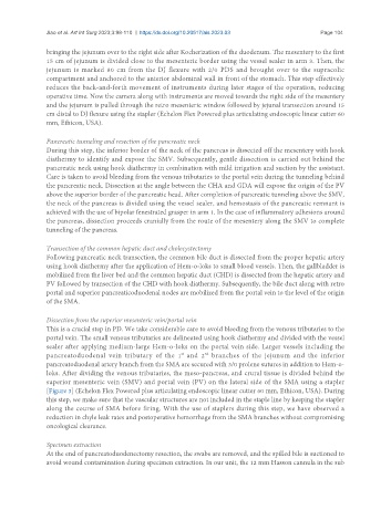
Dissection from the superior mesenteric vein/portal vein
This is a crucial step in PD. We take considerable care to avoid bleeding from the venous tributaries to the
portal vein. The small venous tributaries are delineated using hook diathermy and divided with the vessel
sealer after applying medium-large Hem-o-loks on the portal vein side. Larger vessels including the
pancreatoduodenal vein tributary of the 1 and 2 branches of the jejunum and the inferior
st
nd
pancreatoduodenal artery branch from the SMA are secured with 3/0 prolene sutures in addition to Hem-o-
loks. After dividing the venous tributaries, the meso-pancreas, and crural tissue is divided behind the
superior mesenteric vein (SMV) and portal vein (PV) on the lateral side of the SMA using a stapler
[Figure 3] (Echelon Flex Powered plus articulating endoscopic linear cutter 60 mm, Ethicon, USA). During
this step, we make sure that the vascular structures are not included in the staple line by keeping the stapler
along the course of SMA before firing. With the use of staplers during this step, we have observed a
reduction in chyle leak rates and postoperative hemorrhage from the SMA branches without compromising
oncological clearance.
Specimen extraction
At the end of pancreatoduodenectomy resection, the swabs are removed, and the spilled bile is suctioned to
avoid wound contamination during specimen extraction. In our unit, the 12 mm Hasson cannula in the sub