
Page 110 - Read Online
P. 110
Jiao et al. Art Int Surg 2023;3:98-110 https://dx.doi.org/10.20517/ais.2023.03 Page 102
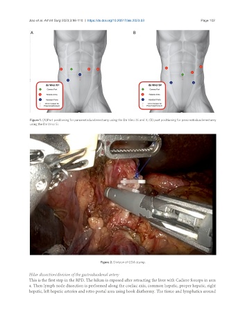
Figure 1. (A)Port positioning for pancreatoduodenectomy using the Da Vinci Xi and X; (B) port positioning for pancreatoduodenectomy
using the Da Vinci Si.
Figure 2. Division of GDA stump.
Hilar dissection/division of the gastroduodenal artery
This is the first step in the RPD. The hilum is exposed after retracting the liver with Cadiere forceps in arm
4. Then lymph node dissection is performed along the coeliac axis, common hepatic, proper hepatic, right
hepatic, left hepatic arteries and retro portal area using hook diathermy. The tissue and lymphatics around