
Page 111 - Read Online
P. 111
Page 103 Jiao et al. Art Int Surg 2023;3:98-110 https://dx.doi.org/10.20517/ais.2023.03
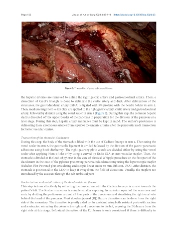
Figure 3. Transection of pancreatic crural tissue.
the hepatic arteries are removed to define the right gastric artery and gastroduodenal artery. Then, a
dissection of Calot’s triangle is done to delineate the cystic artery and duct. After delineation of the
structures, the gastroduodenal artery (GDA) is ligated with 3/0 prolene with the needle holder in arm 3.
Then, medium-large hem-o-lok clips are applied to the right gastric artery, cystic artery and gastroduodenal
artery, followed by division using the vessel sealer in arm 3 [Figure 2]. During this step, the common hepatic
duct is dissected off the upper border of the pancreas in preparation for the division of the pancreas at a
later stage. During this step, hepatic artery anomalies must be kept in mind. The author’s preference is
delineating these anomalous arteries from superior mesenteric arteries after the pancreatic neck transection
for better vascular control.
Transection of the stomach/ duodenum
During this step, the body of the stomach is lifted with the use of Cadiere forceps in arm 4. Then using the
vessel sealer in arm 3, the gastrocolic ligament is divided followed by the division of the gastro-pancreatic
adhesions using hook diathermy. The right gastroepiploic vessels are divided either by using the vessel
sealer after applying Hem-o-loks or by using a curved tip Endo GIA 45 mm vascular stapler. Then, the
stomach is divided at the level of pylorus in the case of classical Whipple procedure or the first part of the
duodenum in the case of the pylorus-preserving pancreatoduodenectomy using the laparoscopic stapler
(Echelon Flex Powered plus articulating endoscopic linear cutter 60 mm, Ethicon, USA). After division, the
stomach is positioned in the LUQ to keep it away from the field of dissection. Usually, the staplers are
introduced by the assistant through the sub-umbilical port.
Kocherization and mobilization of the duodenojejunal flexure
This step is done effectively by retracting the duodenum with the Cadiere forceps in arm 4 towards the
patient’s left. The Kocher maneuver is completed after exposing the anterior aspect of the vena cava and
aorta by dividing the peritoneum around all four parts of the duodenum and visualizing the right renal vein
behind the head of the pancreas. Most duodenojejunal (DJ) flexure dissection can be done from the right
side of the mesentery. The dissection is greatly aided by the assistant using both assistant ports with suction
and a retractor, retracting the colon to the right and duodenum to the left, exposing the DJ flexure from the
right side at this stage. Left-sided dissection of the DJ flexure is only considered if there is difficulty in