
Page 306 - Read Online
P. 306
Page 4 of 19 Cervantes-Gracia et al. Vessel Plus 2020;4:27 I http://dx.doi.org/10.20517/2574-1209.2020.22
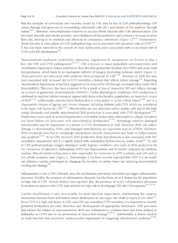
that the interplay of cytotoxicity and viscosity caused by CM, may be key in CIN pathophysiology. CM
causes damage and apoptosis in surrounding endothelial cells (EC) and tubules of the nephron through
iodine [62,63] . Moreover, vasoconstriction is known to increase blood viscosity after CM administration. CM
increased viscosity and tubular pressure, exacerbating renal hypoperfusion and promote a decrease in urine
flow rate, leading to its retention and allowing its continuous cytotoxicity [Figure 1] [64,65] . Furthermore,
blood viscosity is a key player in CVD pathophysiology and is associated with increased risks of CVD [66,67] .
It has also been reported in the context of renal dysfunction and is associated with an increased risk of
[68]
CVD and CKD development .
Vasoconstrictor mediators (endothelin, adenosine, angiotensin II, vasopressin) are known to play a
key role CIN and CVD pathogenesis [65,69-73] . CM is known to cause immediate vasoconstriction and
vasodilation impairment, reduce renal blood flow, decrease glomerular filtration rate (GFR) and cause renal
hypoperfusion, which leads to an inadequate delivery of oxygen, promoting ischemic injury [Figure 1].
These processes are associated with oxidative stress promoted by CM [74,75] . Decrease in GFR has also
[76]
been associated with increased risk in CVD mortality, a feature that reflects kidney damage . Regarding
vasodilatation impairment, it has been suggested to be induced by CM through decreased nitric oxide (NO)
bioavailability. This event has been proposed to be a result of loss of vasoactive NO and cellular damage
on account of generation of peroxynitrite (ONOO ). Under physiological conditions, ROS production is
-
attributed to nephron tubular transport regions with dense mitochondria populations, an important source
of ROS [77,78] . Additionally, mitochondrial dysfunction is a key player in acute kidney injury [79,80] and is a
characteristic feature of ageing, and chronic diseases, including diabetes and CVD, which are considered
to be major risk factors for CIN [81,82] . Mitochondria are also abundant within cardiac cells due to the high
energy demands, and notably mitochondrial ROS production is associated with CVD development [15,83] .
Deleterious events such as arterial hypertension, endothelial dysfunction, atherosclerotic plaque formation
and heart failure are associated with mitochondrial dysfunction [84-86] . Mitophagy removes damaged
mitochondria and its impairment is a feature in CVD development as well. Moreover, ROS can induce
damage in mitochondrial DNA, and damaged mitochondria are important sources of ROS; therefore,
ROS overproduction due to mitophagy impairment disturbs homeostasis and leads to inflammation
and apoptosis [87,88] . As in CIN, excessive ROS production from mitochondria is also associated with NO
[89]
vasodilator impairment and it is tightly linked with endothelial dysfunction in cardiac event . To add
to CIN pathophysiology, oxygen imbalance under hypoxic conditions also leads to ROS production by
the conversion of adenosine triphosphate (ATP) into hypoxanthine and its further reduction by xanthine
oxidase. Mitochondrial dysfunction is also responsible for reduction in ATP synthesis, and will add to
the cellular apoptotic state [Figure 1]. Interestingly, it has been recently reported that CKD in a rat model
can influence cardiac pathologies by changing the function of cardiac tissue and inducing mitochondrial
swelling and damage .
[90]
Inflammation is also a CIN hallmark, since the mechanisms previously described can trigger inflammatory
processes. Notably, the presence of inflammatory elements has also been set as a feature for the population
at high risk of CIN. Several studies have reported that the presence of active inflammatory processes
biomarkers in patients with CVD may attribute its high-risk to developing CIN after CM exposure [30,31,91-94] .
Cardiac insufficiency is also accountable for renal function impairment, emphasizing the complex
interactions between heart and kidney where dysfunction in one organ can result in injury of the other .
[95]
Since CVD is a high-risk factor in CIN, and CIN can exacerbate CVD mortality, it is important to identify
potential biomarkers for early detection and development of appropriate treatments. CIN processes
that induce the release of vasoconstrictors, ROS and inflammatory cytokines have also been defined as
hallmarks in CVDs due to the promotion of myocardial damage [50,96,97] . Additionally, a drastic decline
in renal function may accelerate cardiovascular impairment by triggering inflammatory pathways [95,98] .