
Page 259 - Read Online
P. 259
Idhrees et al. Vessel Plus 2020;4:23 I http://dx.doi.org/10.20517/2574-1209.2020.15 Page 5 of 10
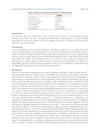
Table 3. Incidence of cardiovascular manifestation in Takasyu’s arteritis
Incidence
Hypertension 34%-79.2%
Pulmonary arteritis 7.1%-18.9%
Coronary artery disease 5.20%-20.1%
Ischemic heart diseae 10.60%
Myocardial failure 6.60%
Aortic regurgitation 33.20%-38.80%
Aortic aneurysm 15.00%-23.30%
Classification
The coronary lesions are classified into 3 types: (1) Type I: stenotic lesion in the ostial segment of the
coronary artery. This is the most commonly encountered type in clinical practice; (2) type II: diffuse
involvement of the coronary arteries or any focal segment of the artery - skip lesions; and (3) type III:
aneurysm of the coronary artery.
Pathogenesis
Active inflammation leads to intimal proliferation and fibrous contraction in the region around the
coronary ostium leading to the narrowing of the coronary artery. Ischemia is one of the major causes of
death in TA patients. Though extremely difficult, a high degree of suspicion is necessary to diagnosis these
patients in the pre-stenotic phase. In patients with diminished/absent pulse, it may take several months/
years, before the coronary artery becomes involved. In a study from Korea, where CT aortogram was
performed in 111 patients, there was a high prevalence of coronary artery abnormalities on coronary CT
angiography, regardless of disease activity or symptoms. Hence, it was recommended to perform coronary
[29]
CT angiography in all patients with TA for additional information .
Management
[30]
Patients on conservative management have a grave prognosis with many dying of cardiac events .
Revascularisation should be considered as early as possible. It is advisable to avoid surgery in the active
phase; however, to avoid any cardiac accidents, revascularisation has to be performed in unstable patients.
In such patients, corticosteroid (CS) or immunosuppressive therapy should be administered at the same
[31]
time to control the inflammatory process . Biologic therapy, with namely anti-IL-6 (tocilizumab, TCZ)
and TNFi (etanercept, infliximab and adalimumab), has been evaluated in TA. Improvement in disease
activity was observed, and TA patients were able to taper or discontinue CSs after initiation of TCZ or
TNFi. Even though there was clinical efficacy, radiological progression was the concern in TA patients on
TCZ. There is evidence favoring the use of TCZ and TNFi in refractory and relapsing TA not responding
to csDMARDs (conventional DMARDs). Abatacept (CTLA4-Ig) use in TA did not meet the primary end-
points in randomized controlled trials (RCTs). Rituximab (anti-CD 20) and ustekinumab (anti-IL-12/23)
in TA gave variable results in isolated case reports [32-34] . High-dose prednisolone (1 mg/kg/day) or its
equivalent is the initial therapy for active TA with tapering dose for 3 to 6 months. CS dose of 0.5 mg/kg/day
together with immunosuppressive agent such as mycophenolate has been recently suggested with equal
[34]
early response. Tapering of steroids can be started in 4 to 6 weeks . Doses below 10 mg/day are associated
with increased relapse . Conventional immunosuppressive agents are started simultaneously with CS or
[34]
while tapering it. There are no RCTs on the use of conventional synthetic disease-modifying antirheumatic
drugs for TA. Weekly methotrexate (15-25 mg/week) therapy has been the first option by many physicians
because of low cost and easy availability. AZA (azathioprine) at 2 mg/kg/day along with CSs for a period
of 12 months showed reduction in inflammatory markers with no radiological progression. Severe TA
(retinal vasculitis, severe AR, myocarditis, or pulmonary vasculitis with or without aneurysm) cases were
given oral cyclophosphamide at 2 mg/kg/day. Mycophenolate mofetil has been a safe and effective drug at
a dose of 2 g/day in treating TA. Both clinical and radiological progression was prevented when given for