
Page 258 - Read Online
P. 258
Page 4 of 10 Idhrees et al. Vessel Plus 2020;4:23 I http://dx.doi.org/10.20517/2574-1209.2020.15
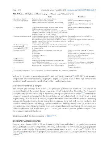
Table 2. Merits and limitations of different imaging modalities to assess Takayasu arteritis
Merits Limitations
Conventional digital Evaluation of severity of stenotic lesions Invasive
subtraction angiography Assment of central blood pressure Risk of contrast induced nephropathy
Concomitant therapeutic intervention as necessary Radiation
Inablity to assess the thickness of arterial
wall
CT angiography Ability to evaluate stenotic and aneurysmal lesions Risk of contrast induced nephropathy
Ability to measure arterial wall thickness Radiation
When used in patients at high suspicion for TA, CTA has a
sensitivity of 95% and specificity of 100%, using catheter-
based angiography as the gold standard
Magnetic resonance imaging Ability to evaluate stenotic and aneurysmal lesions Vessel Decreased sensitivity for smaller branch
wall evaluation (thickening, oedema, degeneration) involvement
Better assement of soft tissue when comapred to CTA No Overestimate degrees of severe stenosis or
radiation exposureSensitivity and specificity of 100% vs occlusion
catheter-based angiography
18F-fluorodeoxyglucose Localise active inflammation and intensity of inflammation Not an angiographic study modality
positron emission Sensitivity and specificity 70.1% and 77.2%for evaluation
tomography (FDG-PET) of disease activity
Duplex ultrasound Ability to evaluate localised areas of stenosis and Unable to provide a “roadmap” of vascular
aneurysm lesions
Non-invasive Unable to perform complete imaging of the
No radiation exposure aortic arch and descending aorta
Nocontrast Operator dependent
Ansthoracic and Non-invasive Unable to provide a ‘roadmap’ of vascular
transesophageal Ability for concomitant assessment of aortic root and lesions
echocardiography aortic valve for insufficiency Unable to differentiate among pathologies
Can be used for surveillance of ascending aorta dilatation, causing hypo echoic aortic wall mural
detection of PHT, and possibly aortic wall thickening thickening
Operator dependent
CT: computed tomography; CTA: CT angiography; TA: Takayasu’s arteritis; PHT: pulmonary hypertension
[10]
and has the potential to assess disease activity and response to treatment . FDG-PET is an operator-
independent, non-invasive metabolic imaging tool helpful in diagnosis of TA. It has a high sensitivity and
specificity, which increases the overall efficacy of the modality in diagnosis.
General consideration in surgery
The disease goes through three phases - pre-pulseless, pulseless and burnt out. This may be an
oversimplification of the complex disease process, and not all patients follow this outline. The few general
principles that physicians should keep in mind when treating such patients are as follows [20-23] : (1) usually an
emergency surgery is not required as the stenotic lesions are well collateralized; (2) it is preferred to avoid
surgery during the active phase. If required, suppress the active disease with medication before considering
surgery; (3) TA patients are often on steroid therapy, making them high-risk surgical candidates due
to effects of medications - for obesity, immunosuppression, bleeding diathesis; and (4) the disease is
progressive, and hence, the patient should be on constant surveillance with medication. It is not uncommon
to see complications such as restenosis, graft occlusion, graft site aneurysm and pseudoaneurysm due to
the progressive nature of the disease.
The incidence of all the disease is shown in Table 3 [6,24,25] .
CORONARY ARTERY DISEASE
Coronary artery disease (CAD) in TA was first described by Frovig and Loken in 1951, and Coronary artery
bypass grafting (CABG) was first performed by Young and colleagues in 1973. Coronary angiographic and
[21]
pathologic studies together have revealed coronary artery lesions in 9 to 11% of cases . CAD in TA is
usually associated with lesions of peripheral branch arteries, and isolated CAD is present in less than 5% of
patients [26-28] .