
Page 257 - Read Online
P. 257
Idhrees et al. Vessel Plus 2020;4:23 I http://dx.doi.org/10.20517/2574-1209.2020.15 Page 3 of 10
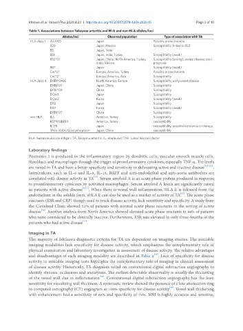
Table 1. Associations between Takayasu arteritis and HLA and non-HLA alleles/loci
Alleles/loci Observed population Type of association with TA
HLA class I A (A10) Japan Possible protective role
B39 Japan, Mexico Susceptibility, linked to B52
B5 Japan, India
B51 Japan, India, Turkey Susceptibility (weak)
B52*01 Japan, China, North America, Turkey, Susceptibility (strong), severe disease, poor
India, Mexico prognosis
B67 Japan Susceptibility (weak)
Cw*07 Europe, America, Turkey Possible protective role
Cw*12 Europe, America, Asia Susceptibility
HLA class II DRB1*0405 North America, Europe Susceptibility, early-onset disease
DRB1*07 Japan, China Susceptibility
DPB1*09 China Susceptibility
DQw1 Japan Susceptibility
DQw2 Korea Susceptibility (weak)
DR2 Japan Susceptibility
DR7 Korea Susceptibility (weak)
DRB1*07 China Susceptibility
non-HLA IL6 America, Turkey Susceptibility
RSP9/LILRB3 America, Turkey susceptibility
IL12B Japan susceptibility, possible resistance to therapy
TNFa 308A/G polymorphism Japan, China susceptibility
HLA: human leukocyte antigen; TA: Takayasu arteritis; IL: interleukin; TNF: tumor necrosis factor
Laboratory findings
Pentraxin 3 is produced in the inflammatory region by dendritic cells, vascular smooth muscle cells,
fibroblasts and macrophages through the trigger of proinflammatory cytokines, especially TNF-α. The levels
are raised in TA and have a better specificity and sensitivity in delineating active and inactive disease [6,11,12] .
Interleukins, such as IL-6 and IL-8, IL-18, BAFF and anti-endothelial and anti-aorta antibodies are
[13]
correlated with disease activity in TA . Serum amyloid A is an acute phase protein produced in response
to proinflammatory cytokines by activated macrophages. Serum amyloid A levels are significantly raised
in patients with active disease [6,14] . When there is vessel wall inflammation, HLA E is released from the
endothelium in the soluble form. sHLA E can also be used as a marker of activity of TA . The acute phase
[15]
reactants (ESR and CRP) though used to track disease activity, lack sensitivity and specificity. A study from
the Cleveland Clinic showed 23% of patients with normal acute phase reactants in the setting of active
[16]
disease . Another analysis from North America showed elevated acute phase reactants in 44% of patients
who were considered to be clinically inactive. Furthermore, ESR was elevated in only three-fourths of the
[17]
patients who had active disease .
Imaging in TA
The majority of Ishikawa diagnostic criteria for TA are dependent on imaging studies. The available
imaging modalities lack specificity for disease activity, which emphasizes the complementary role of
physical examination and laboratory investigation in assessment of disease activity. The relative advantages
[18]
and disadvantages of each imaging modality are described in Table 2 . Lack of specificity for disease
activity in available imaging tests highlights the complementary role of imaging in clinical assessment
of disease activity. Historically, TA diagnosis relied on conventional digital subtraction angiography to
identify stenosis, occlusions and aneurysms. The earliest detectable abnormality is usually the thickening
[10]
of the vessel wall due to inflammation . Conventional digital subtraction angiography has the least
sensitivity for visualizing wall thickness. A systematic review showed the presence of a low attenuation ring
[19]
in computed tomography (CT) angiogram as 100% specificity for disease activity . Vessel wall thickening
with enhancement had a sensitivity of 88% and specificity of 75%. MRI is highly accurate and sensitive,