
Page 14 - Read Online
P. 14
Page 4 of 8 Qin et al. Vessel Plus 2020;4:2 I http://dx.doi.org/10.20517/2574-1209.2019.22
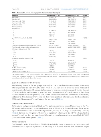
Table 1. Demographic, clinical, and angiographic characteristics of the subjects
Variables Pro-UK group (n = 127) Control group (n = 185) P-value
Age (years) 59.56 ± 12.58 61.34 ± 11.73 0.203
Male [n (%)] 109 (85.83%) 146 (78.92%) 0.121
Hypertension [n (%)] 70 (55.12%) 113 (61.08%) 0.293
Hyperlipemia [n (%)] 34 (26.77%) 69 (37.30%) 0.052
Diabetes [n (%)] 25 (19.69%) 39 (21.08%) 0.764
Smoking [n (%)] 51 (40.16%) 65 (35.14%) 0.367
Killip [n (%)] 0.259
I 101 (79.53%) 149 (80.54%)
II 10 (7.87%) 23 (12.43%)
III 12 (9.45%) 10 (5.41%)
IV 4 (3.15%) 3 (1.62%)
pre-PCI TIMI classification [n (%)] < 0.001
0 107 (84.25%) 121 (65.41%)
1 14 (11.02%) 35 (18.92%)
2 6 (4.72%) 29 (15.68%)
Time from symptom onset to balloon dilatation (h) 6.00 (4.50-9.00) 7.00 (5.00-10.00) 0.193
Time from admission to balloon dilatation (min) 65.00 (48.50-85.00) 64.00 (47.00-93.00) 0.202
Infarction-related artery [n (%)] 0.083
LM 2 (1.57%) 4 (2.16%)
LAD 56 (44.09%) 93 (50.27%)
LCX 8 (6.30%) 23 (12.43%)
RCA 61 (48.03%) 65 (35.14%)
Number of implanted stents [n (%)] 0.864
PTCA 7 (5.51%) 8 (4.32%)
1 stent 91 (71.65%) 134 (72.43%)
2 stent 26 (20.47%) 36 (19.46%)
3 stents 3 (2.36%) 7 (3.78%)
Suction catheter [n (%)] 15 (11.81%) 7 (3.78%) 0.007
IABP used [n (%)] 6 (4.72%) 11 (5.95%) 0.640
Temporary pacemaker [n (%)] 23 (18.11%) 27 (14.59%) 0.406
LM: left main stem; LCX: left circumflex artery; RCA: right coronary artery; IABP: intra-aortic balloon pump; PTCA: percutaneous
transluminal coronary angioplasty; PCI: percutaneous coronary intervention; TIMI: thrombolysis in myocardial infarction; Pro-UK:
prourokinase; LAD: left anterior descending branch
Evaluation of clinical effectiveness
The following indexes of the two groups were analyzed: the TIMI classification of the IRA immediately
after surgery and the corrected TIMI frame count (CTFC) were used to assess the blood perfusion of
culprit vessels; whether the ST segment had decreased by more than 50% at 90 min; and whether the peak
of CK-MB and cTnI had decreased by more than 50%. There were no significant differences in the indexes
of color Doppler echocardiography (LVEF, LVEDd, ventricular aneurysm, and ventricular thrombus)
within seven days after surgery (P > 0.05), but there was a significant difference in the TIMI classification,
CTFC, and CK peak value (P < 0.05) between the two groups [Table 2].
Clinical safety assessment
Eight patients had gastrointestinal bleeding. Two patients experienced cerebral hemorrhage in the Pro-
UK group, while 12 patients experienced gastrointestinal bleeding in the control group. There was no
significant difference in the severity of complications (P > 0.05) or the length of hospital stay (P > 0.05)
[Table 3] between the two groups. There was no significant difference in the VT/VF between the two
groups (P > 0.05), but there was a significant difference in the third-degree atrioventricular block (III° AVB)
(P < 0.05) between the two groups [Table 3].
Follow-up results
Cardiovascular Major Adverse Events (MACEs) is a clinically viable technique for accurate, rapid, and
safe evaluation of myocardial perfusion. In this study, MACE events included stent thrombosis, angina