Page 196 - Read Online
P. 196
Tumscitz et al. Vessel Plus 2019;3:20  I  http://dx.doi.org/10.20517/2574-1209.2018.71                                                   Page 5 of 8

                                  A                              B


                                  C                              D


                                  E                              F


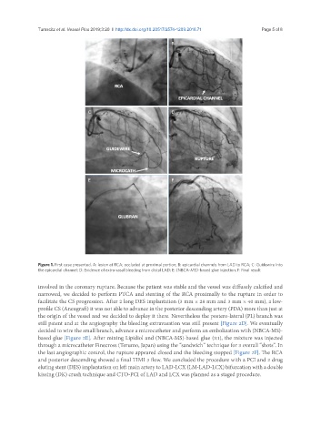
               Figure 1. First case presented. A: lesion at RCA; occluded at proximal portion; B: epicardial channels from LAD to RCA; C: Guidewire into
               the epicardial channel; D: Evidence of extra vasal bleeding from distal LAD; E: (NBCA-MS)-based glue injection; F: Final result

               involved in the coronary rupture. Because the patient was stable and the vessel was diffusely calcified and
               narrowed, we decided to perform PTCA and stenting of the RCA proximally to the rupture in order to
               facilitate the CS progression. After 2 long DES implantation (3 mm × 28 mm and 3 mm × 40 mm), a low-
               profile CS (Aneugraft) it was not able to advance in the posterior descending artery (PDA) more than just at
               the origin of the vessel and we decided to deploy it there. Nevertheless the postero-lateral (PL) branch was
               still patent and at the angiography the bleeding extravasation was still present [Figure 2D]. We eventually
               decided to wire the small branch, advance a microcatheter and perform an embolization with (NBCA-MS)-
               based glue [Figure 2E]. After mixing Lipidiol and (NBCA-MS)-based glue (3:1), the mixture was injected
               through a microcatheter Finecross (Terumo, Japan) using the “sandwich” technique for 3 overall “shots”. In
               the last angiographic control, the rupture appeared closed and the bleeding stopped [Figure 2F]. The RCA
               and posterior descending showed a final TIMI 3 flow. We concluded the procedure with a PCI and 2 drug
               eluting stent (DES) implantation on left main artery to LAD-LCX (LM-LAD-LCX) bifurcation with a double
               kissing (DK) crush technique and CTO-PCI of LAD and LCX was planned as a staged procedure.

