
Page 195 - Read Online
P. 195
Page 4 of 8 Tumscitz et al. Vessel Plus 2019;3:20 I http://dx.doi.org/10.20517/2574-1209.2018.71
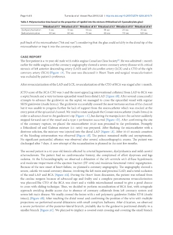
Table 2. Polymerization time based on the proportion of Lipidiol into the mixture (Ethiodized oil: Cyanoakrylate glue)
Ethiodized oil 1:1 Ethiodized oil 2:1 Ethiodized oil 3:1 Ethiodized oil 4:1 Ethiodized oil 5:1 Ethiodized oil 6:1
Start polymerization 5 sec 10 sec 10 sec 18 sec 20 sec 25 sec
End polymerization 40 sec 60 sec 75 sec 85 sec 110 sec 120 sec
pull-back of the microcatheter (“hit and run”) considering that the glue could solidify in the distal tip of the
microcatheter or trap it into the coronary system.
CASE REPORT
The first patient is a 78-year old male with stable angina Canadian Class Society . He was admitted 1 month
[3]
earlier for stable angina and the coronary angiography showed a severe coronary artery disease with critical
stenosis of left anterior descending artery (LAD) and left circumflex artery (LCX) and a CTO of the right
coronary artery (RCA) [Figure 1A]. The case was discussed in Heart Team and surgical revascularization
was excluded by patient’s preference.
After revascularization of the LAD and LCX, revascularization of the CTO of RCA was staged after 1 month.
JCTO score of the RCA CTO was 3 and the most appealing interventional collateral from LAD to RCA was
a septal branch and a very tortuous epicardial vessel from distal LAD [Figure 1B]. After several unsuccessful
attempts to advance the guidewire on the septal, we managed to cross the epicardial vessel with regular
SION guidewire (Asahi Intecc). The guidewire successfully crossed the most tortuous section of the channel
but it was unable to progress further for lack of support from the microcatheter which was stocked at the
entry point of the epicardial channel. We tried to rotate and push the Corsair microcatheter (Asahi Intecc) in
order to advance closer to the guidewire`s tip [Figure 1-C], but during the manipulation the catheter suddenly
stepped forward out of the vessel and a type 3 perforation occurred [Figure 1D]. After confirming the site
of the coronary rupture, we placed the microcatheter 10-20 mm proximal to the perforation. Promptly
a Ethiodized oil and Glubran mixture (1:1 ratio) was prepared. After flushing the microcatheter with a
dextrose solution, the mixture was injected into the distal LAD [Figure 1E]. After 10-15 seconds cessation
of the bleeding extravasation was observed [Figure 1F]. The patient remained stable and asymptomatic.
No significant pericardial effusion was observed after several echocardiographic exams. The patient was
discharged after 7 days. A new attempt of the recanalization is planned in the next few months.
The second patient is a 82-year old female affected by arterial hypertension, dyslipidaemia and mild carotid
atherosclerosis. The patient had no cardiovascular history; she complained of effort dyspnoea and legs
oedema. At the Echocardiography we observed a dilatation of the left ventricle with diffuse hypokinesia
and moderate impairment of the ejection fraction (EF 40%) and moderate functional mitral regurgitation.
Because of the new onset of heart failure, we planned a coronary angiography. The angiography showed a
severe, calcific tri-vessel coronary disease, involving the left main and proximal LAD, and a total occlusion
of the mid LAD and RCA [Figure 2A]. During the Heart Team discussion, the patient was refused from
the cardiac surgeon because of advanced age and frailty and a complete percutaneous revascularization
was planned.The CTO of the RCA was short and a visible microchannel seemed to give a good chance
to cross with sliding technique. Thus, we decided to perform recanalization of RCA first, with antegrade
approach avoiding double access due to absence of coronary collaterals from left coronary system and
severe left main disease. We readly crossed the lesion with a soft polymeric guidewire Fielder XT-R (Asahi
intecc), [Figure 2B]. After reaching the distal vessel and confirming the position of the wire with multiple
projections we performed several dilatations with small compliant balloons. After dilatation, we observed
a severe perforation of the posterior-lateral branch, probably due to the guidewire positioned distally in a
smaller branch [Figure 2C]. We planned to implant a covered stent crossing and covering the small branch