
Page 71 - Read Online
P. 71
Page 2 of 5 Singh et al. Vessel Plus 2018;2:10 I http://dx.doi.org/10.20517/2574-1209.2018.18
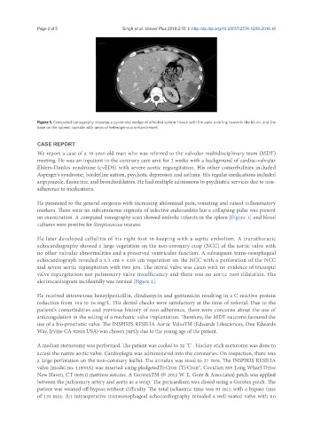
Figure 1. Computed tomography showing a pyramidal wedge of affected splenic tissue with the apex pointing towards the hilum, and the
base on the splenic capsule with areas of heterogenous enhancement
CASE REPORT
We report a case of a 19-year-old man who was referred to the valvular multidisciplinary team (MDT)
meeting. He was an inpatient in the coronary care unit for 2 weeks with a background of cardiac-valvular
Ehlers-Danlos syndrome (cvEDS) with severe aortic regurgitation. His other comorbidities included
Asperger’s syndrome, borderline autism, psychotic depression and asthma. His regular medications included
aripiprazole, fluoxetine, and bronchodilators. He had multiple admissions to psychiatric services due to non-
adherence to medications.
He presented to the general surgeons with increasing abdominal pain, vomiting and raised inflammatory
markers. There were no subcutaneous stigmata of infective endocarditis but a collapsing pulse was present
on examination. A computed tomography scan showed embolic infarcts in the spleen [Figure 1] and blood
cultures were positive for Streptococcus mutans.
He later developed cellulitis of his right foot in keeping with a septic embolism. A transthoracic
echocardiography showed a large vegetation on the non-coronary cusp (NCC) of the aortic valve with
no other valvular abnormalities and a preserved ventricular function. A subsequent trans-oesophageal
echocardiograph revealed a 1.3 cm × 1.05 cm vegetation on the NCC with a perforation of the NCC
and severe aortic regurgitation with two jets. The mitral valve was clean with no evidence of tricuspid
valve regurgitation nor pulmonary valve insufficiency and there was no aortic root dilatation. His
electrocardiogram incidentally was normal [Figure 2].
He received intravenous benzylpenicillin, clindamycin and gentamicin resulting in a C-reactive protein
reduction from 134 to 26 mg/L. His dental checks were satisfactory at the time of referral. Due to the
patient’s comorbidities and previous history of non-adherence, there were concerns about the use of
anticoagulation in the setting of a mechanic valve implantation. Therefore, the MDT outcome favoured the
use of a bio-prosthetic valve. The INSPIRIS RESILIA Aortic ValveTM (Edwards Lifesciences, One Edwards
Way, Irvine CA 92614 USA) was chosen partly due to the young age of the patient.
A median sternotomy was performed. The patient was cooled to 32 ℃ . Hockey stick aortotomy was done to
access the native aortic valve. Cardioplegia was administered into the coronaries. On inspection, there was
a large perforation on the non-coronary leaflet. The annulus was sized to 27 mm. The INSPIRIS RESILIA
valve (model no. 11500A) was inserted using pledgetedTi-Cron (Ti-Cron™, Covidien 555 Long Wharf Drive
New Haven, CT 06511) mattress sutures. A GoretexTM (© 2012 W. L. Gore & Associates) patch was applied
between the pulmonary artery and aorta as a wrap. The pericardium was closed using a Goretex patch. The
patient was weaned off bypass without difficulty. The total ischaemic time was 91 min with a bypass time
of 110 min. An intraoperative transoesophageal echocardiography revealed a well-seated valve with no