
Page 67 - Read Online
P. 67
Page 2 of 4 Shaikhrezai et al. Vessel Plus 2018;2:9 I http://dx.doi.org/10.20517/2574-1209.2018.17
A B C
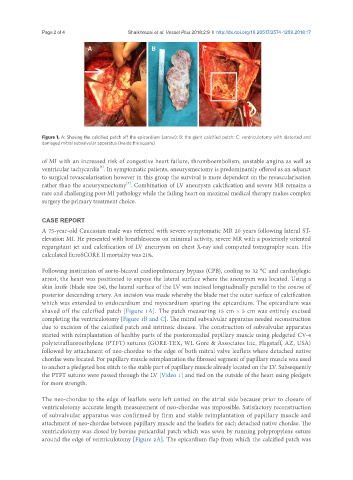
Figure 1. A: Shaving the calcified patch off the epicardium (arrow); B: the giant calcified patch; C: ventriculotomy with distorted and
damaged mitral subvalvular apparatus (inside the square)
of MI with an increased risk of congestive heart failure, thromboembolism, unstable angina as well as
[3]
ventricular tachycardia . In symptomatic patients, aneurysmectomy is predominantly offered as an adjunct
to surgical revascularisation however in this group the survival is more dependent on the revascularisation
[3]
rather than the aneurysmectomy . Combination of LV aneurysm calcification and severe MR remains a
rare and challenging post-MI pathology while the failing heart on maximal medical therapy makes complex
surgery the primary treatment choice.
CASE REPORT
A 75-year-old Caucasian male was referred with severe symptomatic MR 20 years following lateral ST-
elevation MI. He presented with breathlessness on minimal activity, severe MR with a posteriorly oriented
regurgitant jet and calcification of LV aneurysm on chest X-ray and computed tomography scan. His
calculated EuroSCORE II mortality was 21%.
Following institution of aorto-bicaval cardiopulmonary bypass (CPB), cooling to 32 °C and cardioplegic
arrest, the heart was positioned to expose the lateral surface where the aneurysm was located. Using a
skin knife (blade size 24), the lateral surface of the LV was incised longitudinally parallel to the course of
posterior descending artery. An incision was made whereby the blade met the outer surface of calcification
which was extended to endocardium and myocardium sparing the epicardium. The epicardium was
shaved off the calcified patch [Figure 1A]. The patch measuring 15 cm × 5 cm was entirely excised
completing the ventriculotomy [Figure 1B and C]. The mitral subvalvular apparatus needed reconstruction
due to excision of the calcified patch and intrinsic disease. The construction of subvalvular apparatus
started with reimplantation of healthy parts of the posteromedial papillary muscle using pledgeted CV-4
polytetrafluoroethylene (PTFT) sutures (GORE-TEX, WL Gore & Associates Inc, Flagstaff, AZ, USA)
followed by attachment of neo-chordae to the edge of both mitral valve leaflets where detached native
chordae were located. For papillary muscle reimplantation the fibrosed segment of papillary muscle was used
to anchor a pledgeted box stitch to the stable part of papillary muscle already located on the LV. Subsequently
the PTFT sutures were passed through the LV [Video 1] and tied on the outside of the heart using pledgets
for more strength.
The neo-chordae to the edge of leaflets were left untied on the atrial side because prior to closure of
ventriculotomy accurate length measurement of neo-chordae was impossible. Satisfactory reconstruction
of subvalvular apparatus was confirmed by firm and stable reimplantation of papillary muscle and
attachment of neo-chordae between papillary muscle and the leaflets for each detached native chordae. The
ventriculotomy was closed by bovine pericardial patch which was sewn by running polypropylene suture
around the edge of ventriculotomy [Figure 2A]. The epicardium flap from which the calcified patch was