
Page 76 - Read Online
P. 76
Page 2 of 4 Candelaresi et al. Vessel Plus 2018;2:11 I http://dx.doi.org/10.20517/2574-1209.2018.09
A B C D
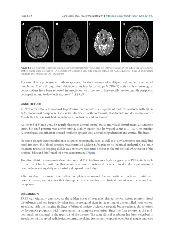
Figure 1. Brain magnetic resonance imaging scans demonstrating: symmetrical high intensity signals in the subcortical white matter
of the occipital lobes at onset on FLAIR images (A), whereas a very mild changes on DWI and ADC sequences (B and C), and imaging
remission after 10 days on FLAIR images (D)
Bortezomib is a proteasome inhibitor approved for the treatment of multiple myeloma and mantle cell
lymphoma. It acts through the inhibition of nuclear factor kappa B (NF-kB) activity. Few neurological
complications have been reported in association with the use of bortezomib, predominantly peripheral
[2-7]
neuropathies, and to date, only six cases of PRES.
CASE REPORT
In November 2016, a 72-year-old hypertensive man received a diagnosis of multiple myeloma with IgGk/
IgAλ monoclonal component. He was initially treated with bortezomib, thalidomide and dexamethasone. In
March 2017, he was switched on melphalan, prednisone and bortezomib.
At the end of March 2017, he acutely developed altered mental status and visual disturbances. At symptom
onset, his blood pressure was 170/80 mmHg, slightly higher than his typical values (140-150/70-80 mmHg).
A neurological examination showed nonfluent aphasia with altered comprehension and cortical blindness.
No acute changes were revealed on a computed tomography scan, as well as in any laboratory test, including
renal function. His blood pressure was controlled adding nifedipine to his habitual enalapril. On a brain
magnetic resonance imaging (MRI) scan extensive vasogenic oedema in the subcortical white matter of the
occipital lobes and left frontal lobe was demonstrated [Figure 1].
The clinical history, neurological examination and MRI findings were highly suggestive of PRES, attributable
to the use of bortezomib. Further administration of bortezomib was withheld and a short course of
dexamethasone 8 mg daily was started and tapered over 5 days.
After 10 days from onset, the patient completely recovered. He was switched on lenalidomide and
dexamethasone, and at 6-month follow-up he is experiencing a serological remission of the monoclonal
component.
DISCUSSION
PRES was originally described as the sudden onset of headache, altered mental status, seizures, visual
disturbances and less frequently other focal neurological signs in the setting of uncontrolled hypertension,
associated with the imaging findings of bilateral parieto-occipital vasogenic brain oedema, characterized
by favourable prognosis with improvement or complete resolution. Since the first reports in the mid-
90s, much has changed in the spectrum of the disease. The same clinical syndrome has been described in
association with atypical radiological patterns, involving frontal and temporal lobes, basal ganglia and even