
Page 182 - Read Online
P. 182
Page 8 of 13 Misra et al. Vessel Plus 2022;6:32 https://dx.doi.org/10.20517/2574-1209.2021.104
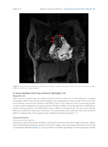
Figure 5. Late gadolinium enhancement (LGE) sequence image showing dilated right ventricle, and late enhancement in right
ventricular outflow tract (red arrowheads).
CT SCAN IMAGING FOR EVALUATION OF REPAIRED TOF
Diagnostic role
Improvement in technology and significant advancement in the area of multi-detector computed
tomography (MDCT) has revolutionized imaging in the congenital heart disease world. TOF is one of the
most common cyanotic heart diseases, and MDCT plays a very important role in answering specific
questions in the management of repaired patients with this condition. Foremost indication would include
patients with pacemakers and defibrillators where CMR is contraindicated. The next most relevant
indication would be for anatomical assessment, 3D reconstruction and/or printing. With some advanced
MDCT, volumetric data can be acquired using a single breath-hold examination [30,31,32] .
Clinical information
Pulmonary arterial anatomy:
Information obtained from this modality would include anatomic detail about right ventricular outflow
tract/conduit/branch pulmonary arteries in terms of its origin, course, narrowing, obstruction, stenosis, and
or aneurysmal dilatation [Figure 6]. It may provide an excellent opportunity to assess pulmonary arterial