
Page 181 - Read Online
P. 181
Misra et al. Vessel Plus 2022;6:32 https://dx.doi.org/10.20517/2574-1209.2021.104 Page 7 of 13
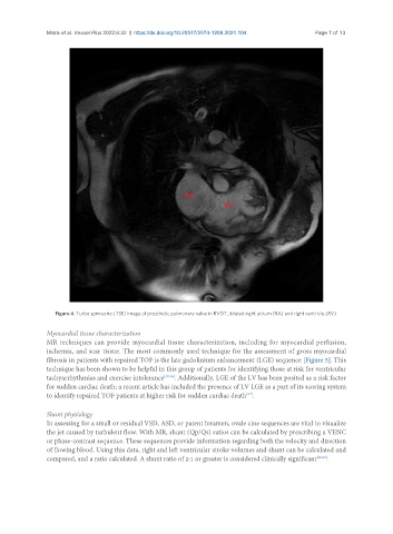
Figure 4. Turbo spin-echo (TSE) image of prosthetic pulmonary valve in RVOT, dilated right atrium (RA) and right ventricle (RV).
Myocardial tissue characterization
MR techniques can provide myocardial tissue characterization, including for myocardial perfusion,
ischemia, and scar tissue. The most commonly used technique for the assessment of gross myocardial
fibrosis in patients with repaired TOF is the late gadolinium enhancement (LGE) sequence [Figure 5]. This
technique has been shown to be helpful in this group of patients for identifying those at risk for ventricular
tachyarrhythmias and exercise intolerance [25,26] . Additionally, LGE of the LV has been posited as a risk factor
for sudden cardiac death; a recent article has included the presence of LV LGE as a part of its scoring system
[27]
to identify repaired TOF patients at higher risk for sudden cardiac death .
Shunt physiology
In assessing for a small or residual VSD, ASD, or patent foramen, ovale cine sequences are vital to visualize
the jet caused by turbulent flow. With MR, shunt (Qp/Qs) ratios can be calculated by prescribing a VENC
or phase-contrast sequence. These sequences provide information regarding both the velocity and direction
of flowing blood. Using this data, right and left ventricular stroke volumes and shunt can be calculated and
compared, and a ratio calculated. A shunt ratio of 2:1 or greater is considered clinically significant [28,29] .