
Page 60 - Read Online
P. 60
Page 8 of 10 Licordari et al. Vessel Plus 2022;6:12 https://dx.doi.org/10.20517/2574-1209.2021.86
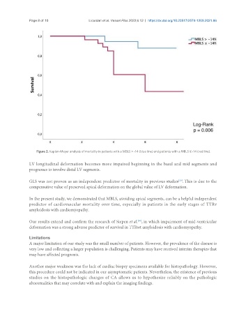
Figure 2. Kaplan-Meyer analysis of mortality in patients with a MBLS > -14 (blue line) and patients with a MBLS ≤ -14 (red line).
LV longitudinal deformation becomes more impaired beginning in the basal and mid segments and
progresses to involve distal LV segments.
[20]
GLS was not proven as an independent predictor of mortality in previous studies . This is due to the
compensative value of preserved apical deformation on the global value of LV deformation.
In the present study, we demonstrated that MBLS, avoiding apical segments, can be a helpful independent
predictor of cardiovascular mortality over time, especially in patients in the early stages of TTRv
amyloidosis with cardiomyopathy.
Our results extend and confirm the research of Siepen et al. , in which impairment of mid-ventricular
[20]
deformation was a strong adverse predictor of survival in TTRwt amyloidosis with cardiomyopathy.
Limitations
A major limitation of our study was the small number of patients. However, the prevalence of the disease is
very low and collecting a larger population is challenging. Patients may have received interim therapies that
may have affected prognosis.
Another major weakness was the lack of cardiac biopsy specimens available for histopathology. However,
this procedure could not be indicated in our asymptomatic patients. Nevertheless, the existence of previous
studies on the histopathologic changes of CA allows us to hypothesize reliably on the pathologic
abnormalities that may correlate with and explain the imaging findings.