
Page 59 - Read Online
P. 59
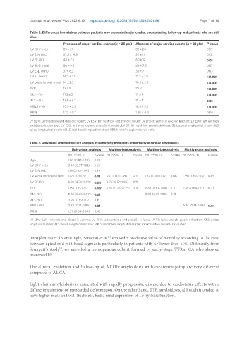
Licordari et al. Vessel Plus 2022;6:12  https://dx.doi.org/10.20517/2574-1209.2021.86  Page 7 of 10

               Table 2. Differences in variables between patients who presented major cardiac events during follow-up and patients who are still
               alive
                               Presence of major cardiac events (n = 25 pts) Absence of major cardiac events (n = 21 pts) P-value
                LV EDV (mL)    85 ± 21                            82 ± 20                           0.57
                LV ESV (mL)    37.5 ± 14.5                        32 ± 11                           0.13
                LV EF (%)      48 ± 7.5                           60 ± 12                           0.01
                LV EDD (mm)    50 ± 9.5                           49 ± 7.5                          0.57
                LV ESD (mm)    31 ± 8.3                           30 ± 7                            0.63
                LV ST (mm)     16.2 ± 2.8                         12.3 ± 2.5                        < 0.001
                LV posterior wall (mm) 14 ± 2.5                   10.5 ± 2.5                        < 0.001
                E/E’           13 ± 5                             7 ± 4                             < 0.001
                GLS (-%)       11.5 ± 3                           19 ± 4                            < 0.001
                ALS (-%)       13.5 ± 6.7                         18 ± 4                            0.01
                MBLS (-%)      10.5 ± 2.5                         16.5 ± 5.3                        < 0.001
                RRSR           1.32 ± 0.7                         1.20 ± 0.4                        0.50

               LV EDV: Left ventricle end diastolic volum; LV ESV: left ventricle end systolic volum; LV EF: left ventricle ejection fraction; LV EDD: left ventricle
               end diastolic diameter; LV ESD: left ventricle end diastolic diameter; LV ST: left ventricle septal thickness; GLS: global longitudinal strain; ALS:
               apical longitudinal strain; MBLS: mid-basal longitudinal strain; RRSR: relative regional strain ratio.


               Table 3. Univariate and multivariate analysis in identifying predictors of mortality in cardiac amyloidosis
                                   Univariate analysis  Multivariate analysis  Multivariate analysis  Multivariate analysis
                                  HR (95%CI)  P-value HR (95%CI)  P-value HR (95%CI)  P-value HR (95%CI)  P-value
                Age               1.02 (0.97-1.08)  0.29
                LV EDV (mL)       0.99 (0.97-1.01)  0.35
                LV ESV (mL)       1.01 (0.98-1.04)  0.39
                LV septal thickness (mm)  1.17 (1.03-1.32)  0.01  0.12 (0.01-1.89)  0.13  1.37 (1.00-1.87)  0.06  1.39 (0.95-2.05)  0.09
                LV EF (%)         0.83 (0.70-0.99)  0.04  0.76 (0.54-1.06)  0.11
                E/E’              1.15 (1.06-1.25)  0.001  6.30 (0.71-55.35)  0.10  0.83 (0.67-1.04)  0.11  0.85 (0.64-1.13)  0.27
                GLS (%)           0.86 (0.76-0.97)  0.01              0.88 (0.73-1.06)  0.18
                ALS (%)           0.96 (0.89-1.02)  0.15
                MBLS (%)          0.85 (0.71-0.96)  0.01                                0.84 (0.71-0.98)  0.04
                RRSR              1.37 (0.64-2.99)  0.42
               LV EDV: Left ventricle end diastolic volume; LV ESV: left ventricle end systolic volume; LV EF: left ventricle ejection fraction; GLS: global
               longitudinal strain; ALS: apical longitudinal strain; MBLS: mid-basal longitudinal strain; RRSR: relative regional strain ratio.

               transplantation. Interestingly, Senapati et al.  showed a predictive value of mortality according to the ratio
                                                     [9]
               between apical and mid-basal segments particularly in patients with EF lower than 45%. Differently from
               Senapati’s study , we enrolled a homogenous cohort formed by early-stage TTRm CA who showed
                             [9]
               preserved EF.

               The clinical evolution and follow-up of ATTRv amyloidosis with cardiomyopathy are very different
               compared to AL CA.


               Light-chain amyloidosis is associated with rapidly progressive disease due to cardiotoxic effects with a
               diffuse impairment of myocardial deformation. On the other hand, TTR amyloidosis, although it tended to
               have higher mass and wall thickness, had a mild depression of LV systolic function.