Page 132 - Read Online
P. 132

De Gaspari et al. Vessel Plus 2022;6:57    https://dx.doi.org/10.20517/2574-1209.2022.05                                                                                       Page 7 of 13


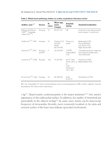
                          Table 2. Whole heart-pathology studies on cardiac amyloidosis: literature review

                                                                 Mean age,
                          Author, year  ref    Source    N.      years         Amyloid        Amyloid localization        Pattern of          Valvular         Vascular              Amyloid           Clinical
                                                         cases                 typing                                     infiltration        involvement      involvement           quantification    correlation
                                                                 (range)

                          Roberts and Waller,   Autopsy  54      64 (21-97)    ND             Interstitium and endocardium  ND                At least 1 valve   No involvement in   ND                Hemodynamic
                               [39]
                          1983    (including                                                  in all; always 1 or both atria                  involved in all  epicardial coronary                     in 16%
                                    [38]
                          cases from   )                                                                                                                       artery                                  Echo in 20%
                                                                                                                                                               Intramural vessels
                                                                                                                                                               involved in all
                                    [40]
                          Smith et al.  , 1984  Autopsy  47      Primary 57.6  Primary vs.    Endocardial 70%             Nodular 49%         Mitral 60%       Present in 19 primary   ND              ND
                                                                 (35-83)       senile         Pericardial 36%             Perifiber 28%       Aortic 23%       and 1 senile (total
                                                                 Senile                       Left atrium 91%             Mixed 17%           Tricuspid 50%    43%)
                                                                 83 (70-89)                   Right atrium 81%                                Pulmonary 30%
                                    [41]
                          Leone et al.  , 2012  Autopsy   9      56 (54-60)    5 ATTR         Computed analysis: trabecular  Diffuse 55.6%    ND               ND                    Quantitative (%)  Echo in all
                                               and HT                          (unspecified)   and subendocardial layers   Segmental 22.2%                                                             CMR in 11%
                                                                               4 AL           most infiltrated            Subendocardial
                                                                                                                          22.2%
                                     [42]
                          Larsen et al.  , 2016 Autopsy  108     75 (31-89)    60 ATTRwt      Interstitium 85%            Pericellular 66%    ND               Epicardial arteries 0%   Semi-quantitative  ND
                                                                               44 AL (32 λ, 12  Endocardium 41%           Nodular 28%                          Epicardial vasa
                                                                               κ)                                                                              vasorum 18%
                                                                               2 AA                                                                            Epicardial veins 48%
                                                                               1 AApoAIV                                                                       Intramyocardial
                                                                               1 AL + AH                                                                       arteries 72%
                                                                                                                                                               Intramyocardial veins
                                                                                                                                                               9%
                                     [43]
                          Porcari et al.  , 2021 Autopsy  24     86 (84-91)    12 AL          Interstitium in 75%         LV non-diffuse 65%  ND               Vascular only in 25%  Semi-quantitative  Echo in 67%
                                                                               12 ATTR                                    vs. diffuse 35%

                          ND: Not disposable; HT: heart transplantation; echo: echocardiography; CMR: cardiac magnetic resonance; ATTR: transthyretin amyloid; AL: light chain amyloidosis; AA: serum amyloid A, apolipoprotein A-IV
                          amyloidosis; AH: heavy chain amyloidosis.


                               [45]
                          1 kg . Hypertrophic cardiomyopathy is the major mimicker                   [46,47] , but amyloid hearts are usually firmer with additional atrial wall thickening and rugged
                          appearance of the endocardial surface. In addition, the marker of interatrial septum thickening can help to distinguish CA from hypertrophic cardiomyopathy,
                          particularly in the clinical setting . In some cases, hearts can be macroscopically normal                    [39,40] . CA, in particular the AL form, is also associated with high
                                                                   [48]
                          frequency of intracardiac thrombi, most commonly localized in the atria and linked to the worst prognosis                          [39,49] . Rough or irregular whitish patches on the
                          external surface of the heart may indicate epicardial involvement.
   127   128   129   130   131   132   133   134   135   136   137