
Page 131 - Read Online
P. 131
Page 6 of 13 De Gaspari et al. Vessel Plus 2022;6:57 https://dx.doi.org/10.20517/2574-1209.2022.05
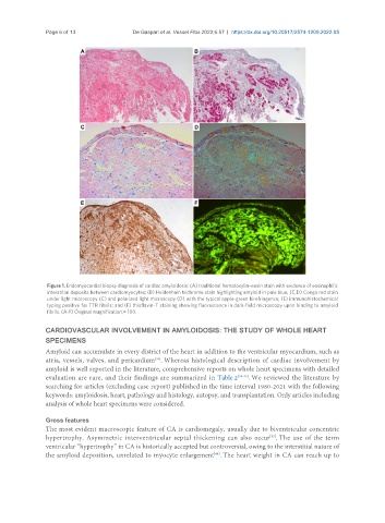
Figure 1. Endomyocardial biopsy diagnosis of cardiac amyloidosis: (A) traditional hematoxylin–eosin stain with evidence of eosinophilic
interstitial deposits between cardiomyocytes; (B) Heidenhain trichrome stain highlighting amyloid in pale blue; (C,D) Congo red stain
under light microscopy (C) and polarized light microscopy (D) with the typical apple-green birefringence; (E) immunohistochemical
typing positive for TTR fibrils; and (F) thioflavin-T staining showing fluorescence in dark-field microscopy upon binding to amyloid
fibrils. (A-F) Original magnification,× 100.
CARDIOVASCULAR INVOLVEMENT IN AMYLOIDOSIS: THE STUDY OF WHOLE HEART
SPECIMENS
Amyloid can accumulate in every district of the heart in addition to the ventricular myocardium, such as
atria, vessels, valves, and pericardium . Whereas histological description of cardiac involvement by
[35]
amyloid is well reported in the literature, comprehensive reports on whole heart specimens with detailed
evaluation are rare, and their findings are summarized in Table 2 [38-43] . We reviewed the literature by
searching for articles (excluding case report) published in the time interval 1980-2021 with the following
keywords: amyloidosis, heart, pathology and histology, autopsy, and transplantation. Only articles including
analysis of whole heart specimens were considered.
Gross features
The most evident macroscopic feature of CA is cardiomegaly, usually due to biventricular concentric
[35]
hypertrophy. Asymmetric interventricular septal thickening can also occur . The use of the term
ventricular “hypertrophy” in CA is historically accepted but controversial, owing to the interstitial nature of
the amyloid deposition, unrelated to myocyte enlargement . The heart weight in CA can reach up to
[44]