


Page 43 - Read Online
P. 43
Raffetto et al. Vessel Plus 2021;5:36  https://dx.doi.org/10.20517/2574-1209.2021.16  Page 9 of 30


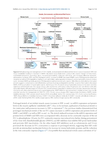
                Figure 2. Pathophysiology and management of CVD. Genetic, environmental and behavioral risk factors cause an increase in HIFs and
                tissue metabolites leading to increases in MMPs. Increased venous hydrostatic pressure also causes changes in shear stress,
                endothelial dysfunction, glycocalyx injury, increased permeability, leukocyte infiltration, and increased adhesion molecules,
                inflammatory cytokines, ROS, and RNS, leading to further increases in MMPs. Increased MMPs cause VSM hyperpolarization and
                relaxation as well as ECM degradation leading to vein wall dilation, and progressive increases in venous hydrostatic pressure (vicious
                cycle). Increased MMPs generally promote ECM degradation particularly in atrophic regions. Other theories (indicated by interrupted
                arrows) suggest a compensatory anti-inflammatory pathway involving prostaglandins and their receptors that leads to decreased
                MMPs, ECM accumulation in hypertrophic regions, and tortuous VVs. Increased venous hydrostatic pressure in the lower extremity
                saphenous and femoral veins also causes venous valve dysfunction and venous reflux. Progressive vein wall dilation and valve
                dysfunction lead to different stages of CVD and CVI. Current treatment (presented in shaded arrows) includes compression stockings,
                venotonics and other pharmacological and surgical approaches. MMP inhibitors (also presented in shaded arrows) may provide
                potential tools for the management of CVD/CVI. CVD: Chronic venous disease; HIFs: hypoxia inducible factors; MMPs: matrix
                metalloproteinases; VSM: vascular smooth muscle; ECM: extracellular matrix; VVs: varicose veins; CVI: chronic venous insufficiency;
                TIMPs: tissue inhibitors of metalloproteinases; ICAM-1: intercellular adhesion molecule-1; VCAM-1: vascular cell adhesion molecule-1;
                siRNA: small interfering RNA; ROS: reactive oxygen species; RNS: reactive nitrogen species; SDX: sulodexide.

               Prolonged stretch of rat skeletal muscle causes increases in HIF-1α and -2α mRNA expression and protein
               levels in the muscle capillaries’ endothelial cells . Also, in the rat heart, application of mechanical stretch to
                                                       [67]
               the ventricular wall promotes increases in HIF-1α expression . Our previous studies demonstrated that
                                                                    [68]
               prolonged mechanical stretch of the rat IVC causes increases in mRNA expression and protein levels of
               MMP-2 and MMP-9 as well as HIF-1α and -2α. The stretch-induced increases in the mRNA expression and
               protein levels of MMPs and HIFs were accompanied with a decrease in the contractile response of the rat
               IVC to phenylephrine. Of note, the IVC contractile response was reduced even further during pretreatment
               of the veins with dimethyloxallyl glycine (DMOG), an HIF stabilizer that inhibits HIF-prolyl hydroxylase
               and prevents HIF inactivation. On the other hand, HIF inhibitors such as echinomycin and U0126
               prevented the decrease in IVC contraction in response to prolonged stretch, suggesting that HIF could
               provide an intermediary mechanism between the increase in venous hydrostatic pressure and the reduction
               in the vein contractile response [Figure 3] . It is possible that mechanical stretch causes activation of Ca
                                                                                                         2+
                                                   [66]

