


Page 40 - Read Online
P. 40
Page 6 of 30                  Raffetto et al. Vessel Plus 2021;5:36  https://dx.doi.org/10.20517/2574-1209.2021.16

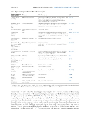
               Table 1. Representative genetic factors in VVs and venous leg ulcer
                Gene chromosome   Disease           Manifestations                          Ref.
                locus, variants
                COL3A1, COL5A1,   Ehlers-Danlos syndrome  Connective tissue disorders, abnormal collagen synthesis, joint  [39,224]
                ADAMTS2                             hypermobility, distensible skin, ocular disease, bone
                                                    deformities and fragility, vascular and visceral tissue wall
                                                    fragility and susceptibility to rupture, VVs
                von-Hippel Lindau   Chuvash polycythemia  Defective oxygen sensing, increased HIF-1α, increased serum   [225-227]
                gene mutation                       erythropoietin and hemoglobin, VVs, tendency for vertebral
                3p25                                hemangiomas, thrombosis, bleeding, and stroke
                598>T
                G6PC3 gene mutation  Severe congenital neutropenia   VVs, venous leg ulcer  [228]
                               type 4
                Desmuslin gene   VVs                Decreased intermediate filament protein desmuslin in VSM,   [48,117,120,229,230]
                deficiency                          smooth muscle switch from contractile to synthetic phenotype,
                15q26.3                             weakening of vein wall, venous dilation. Increased MMP-2 and
                                                    collagen
                Thrombomodulin (-  Deep venous thrombosis, VVs  Dysregulation of thrombin, thrombus formation  [231]
                1208/-1209 TT
                deletion)
                Translocation of   Klippel-Trenaunay syndrome  Cutaneous capillary          [232,233]
                chromosome 8q22.3                   malformations (port wine stain), VVs, bone and soft tissues
                & 14q13                             hypertrophy
                EII3K or VG5Q gene
                mutation on
                chromosome 5
                FOXC2 gene mutation  Lymphoedema distichiasis  Lymphedema, distichiasis (extra eyelashes from meibomian   [234-236]
                16q24.3                             glands), VVs, congenital heart defects, vertebral anomalies,
                                                    extradural cysts, ptosis, cleft palate
                Notch3 gene mutation  CADASIL       Cerebral autosomal-dominant arteriopathy with subcortical   [46]
                1279G>T                             infarcts and leukoencephalopathy, VVs
                Trisomies      VVs                  Abnormalities in cell lines from patients with VVs  [237]
                chromosomes 7, 12, 18
                Monosomy
                chromosome 14
                F13A1 gene     Factor XIII deficiency  Delayed venous ulcer healing         [54]
                HFE gene mutation   Increased iron deposition  Venous ulcer exacerbation    [51]
                C282Y and H63D
                MTFR gene      Decreased            Hyperhomocystinemia, VVs, CVI           [238,239]
                SNP C677T      methylenetetrahydrofolate
                               reductase activity
                SLC40A1        Impaired iron metabolism,   CVD, venous ulcer                [240]
                SNP 8CG        increased iron deposition
                MMP-12         Altered MMP activity  Venous ulcer                           [240]
                SNP 82AA
                FGFR-2         mRNA instability, reduced   CVI, non-healing venous ulcer    [241]
                SNP 2451AG     wound healing

               VVs: Varicose veins; VSM: vascular smooth muscle; MMP: matrix metalloproteinases; CADASIL: cerebral autosomal dominant arteriopathy with
               subcortical infarcts and leukoencephalopathy; CVI: chronic venous insufficiency; CVD: chronic venous disease.

               were robustly associated with VVs, including genes encoding for blood pressure, vascular mechanosensing
               channels, vascular maturation, development and integrity, and genes near the hemochromatosis gene that is
               strongly associated with VLU and DVT . Patients with Ehlers-Danlos syndrome show increased
                                                    [38]
               propensity to developing vascular pathologies and VVs . Ehlers-Danlos syndrome includes several
                                                                  [39]
               connective tissue disorders that involve abnormal collagen synthesis and could be manifested in the form of
               distensible skin, joint hypermobility, bone fragility and deformity, ocular disease, and cardiovascular and
               visceral disorders in which the blood vessels and visceral tissue walls become more fragile and prone to
               rupture. Patients with vascular Ehlers-Danlos syndrome have defective COL3A1 gene and are more
               susceptible to vascular disease and VVs [39,40] . Of note, patients with hereditary connective tissue syndromes

