


Page 38 - Read Online
P. 38
Page 4 of 30                  Raffetto et al. Vessel Plus 2021;5:36  https://dx.doi.org/10.20517/2574-1209.2021.16


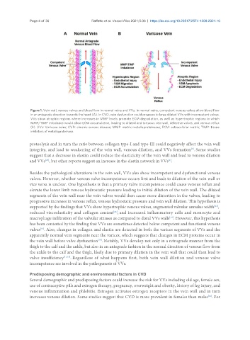
                Figure 1. Vein wall, venous valves and blood flow in normal veins and VVs. In normal veins, competent venous valves allow blood flow
                in an antegrade direction towards the heart (A). In CVD, vein dysfunction could progress to large dilated VVs with incompetent valves.
                VVs show atrophic regions where increases in MMP levels promote ECM degradation, as well as hypertrophic regions in which
                MMP/TIMP imbalance would allow ECM accumulation, leading to dilated and tortuous vein wall, defective valves, and venous reflux
                (B). VVs: Varicose veins; CVD: chronic venous disease; MMP: matrix metalloproteinases; ECM: extracellular matrix; TIMP: tissue
                inhibitors of metalloproteinase.

               proteolysis and in turn the ratio between collagen type-I and type-III could negatively affect the vein wall
               integrity, and lead to weakening of the vein wall, venous dilation, and VVs formation . Some studies
                                                                                            [2]
               suggest that a decrease in elastin could reduce the elasticicity of the vein wall and lead to venous dilation
                       [20]
               and VVs , but other reports suggest an increase in the elastin network in VVs .
                                                                                 [2]
               Besides the pathological alterations in the vein wall, VVs also show incompetent and dysfunctional venous
               valves. However, whether venous valve incompetence occurs first and leads to dilation of the vein wall or
               vice versa is unclear. One hypothesis is that a primary valve incompetence could cause venous reflux and
               elevate the lower limb venous hydrostatic pressure leading to initial dilation of the vein wall. The dilated
               segments of the vein wall near the vein valves would then cause more distortion in the valves, leading to
               progressive increases in venous reflux, venous hydrostatic pressure and vein wall dilation. This hypothesis is
               supported by the findings that VVs show hypertrophic venous valves, augmented valvular annulus width ,
                                                                                                       [21]
                                                       [22]
               reduced viscoelasticity and collagen content , and increased inflammatory cells and monocyte and
               macrophage infiltration of the valvular sinuses as compared to distal VVs walls . However, this hypothesis
                                                                                  [23]
               has been contested by the finding that VVs are sometimes detected below competent and functional venous
               valves . Also, changes in collagen and elastin are detected in both the varices segments of VVs and the
                    [13]
               apparently normal vein segments near the varices, which suggests that changes in ECM proteins occur in
               the vein wall before valve dysfunction . Notably, VVs develop not only in a retrograde manner from the
                                                [16]
               thigh to the calf and the ankle, but also in an antegrade fashion in the normal direction of venous flow from
               the ankle to the calf and the thigh, likely due to primary dilation in the vein wall that could then lead to
               valve insufficiency [11,13] . Regardless of what happens first, both vein wall dilation and venous valve
               incompetence are involved in the pathogenesis of VVs.

               Predisposing demographic and environmental factors in CVD
               Several demographic and predisposing factors could increase the risk for VVs including old age, female sex,
               use of contraceptive pills and estrogen therapy, pregnancy, overweight and obesity, history of leg injury, and
               venous inflammation and phlebitis. Estrogen activates estrogen receptors in the vein wall and in turn
               increases venous dilation. Some studies suggest that CVD is more prevalent in females than males . For
                                                                                                    [12]

