
Page 67 - Read Online
P. 67
Abdel-Halim et al. Vessel Plus 2022;6:8 https://dx.doi.org/10.20517/2574-1209.2021.40 Page 9 of 14
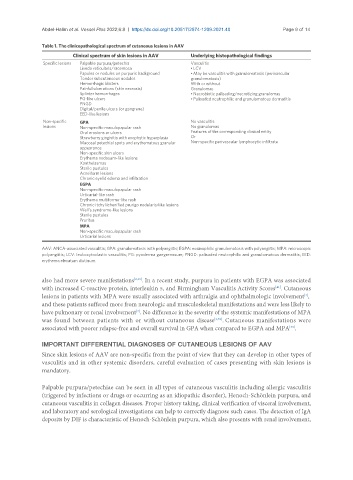
Table 1. The clinicopathological spectrum of cutaneous lesions in AAV
Clinical spectrum of skin lesions in AAV Underlying histopathological findings
Specific lesions Palpable purpura/petechia Vasculitis
Livedo reticularis/racemosa • LCV
Papules or nodules on purpuric background • May be vasculitis with granulomatosis (perivascular
Tender subcutaneous nodules granulomatosis)
Hemorrhagic blisters With or without
Painful ulcerations (skin necrosis) Granulomas
Splinter hemorrhages • Necrobiotic palisading/necrotizing granulomas
PG-like ulcers • Palisaded neutrophilic and granulomatous dermatitis
PNGD
Digital/penile ulcers (or gangrene)
EED-like lesions
Non-specific GPA No vasculitis
lesions Non-specific maculopapular rash No granulomas
Oral erosions or ulcers Features of the corresponding clinical entity
Strawberry gingivitis with exophytic hyperplasia Or
Mucosal petechial spots and erythematous granular Non-specific perivascular lymphocytic infiltrate
appearance
Non-specific skin ulcers
Erythema nodosum-like lesions
Xanthelasmas
Sterile pustules
Acneiform lesions
Chronic eyelid edema and infiltration
EGPA
Non-specific maculopapular rash
Urticarial-like rash
Erythema multiforme-like rash
Chronic itchy lichenified prurigo nodularis-like lesions
Well’s syndrome-like lesions
Sterile pustules
Pruritus
MPA
Non-specific maculopapular rash
Urticarial lesions
AAV: ANCA-associated vasculitis; GPA: granulomatosis with polyangiitis; EGPA: eosinophilic granulomatosis with polyangiitis; MPA: microscopic
polyangiitis; LCV: leukocytoclastic vasculitis; PG: pyoderma gangrenosum; PNGD: palisaded neutrophilic and granulomatous dermatitis; EED:
erythema elevatum diutinum.
also had more severe manifestations [2,39] . In a recent study, purpura in patients with EGPA was associated
[41]
with increased C-reactive protein, interleukin 5, and Birmingham Vasculitis Activity Scores . Cutaneous
lesions in patients with MPA were usually associated with arthralgia and ophthalmologic involvement ,
[4]
and these patients suffered more from neurologic and musculoskeletal manifestations and were less likely to
have pulmonary or renal involvement . No difference in the severity of the systemic manifestations of MPA
[2]
was found between patients with or without cutaneous disease [2,39] . Cutaneous manifestations were
associated with poorer relapse-free and overall survival in GPA when compared to EGPA and MPA .
[39]
IMPORTANT DIFFERENTIAL DIAGNOSES OF CUTANEOUS LESIONS OF AAV
Since skin lesions of AAV are non-specific from the point of view that they can develop in other types of
vasculitis and in other systemic disorders, careful evaluation of cases presenting with skin lesions is
mandatory.
Palpable purpura/petechiae can be seen in all types of cutaneous vasculitis including allergic vasculitis
(triggered by infections or drugs or occurring as an idiopathic disorder), Henoch-Schönlein purpura, and
cutaneous vasculitis in collagen diseases. Proper history taking, clinical verification of visceral involvement,
and laboratory and serological investigations can help to correctly diagnose such cases. The detection of IgA
deposits by DIF is characteristic of Henoch-Schönlein purpura, which also presents with renal involvement,