
Page 63 - Read Online
P. 63
Abdel-Halim et al. Vessel Plus 2022;6:8 https://dx.doi.org/10.20517/2574-1209.2021.40 Page 5 of 14
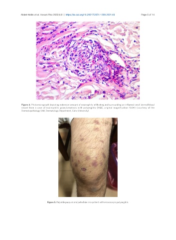
Figure 4. Photomicrograph depicting extensive amount of eosinophils infiltrating and surrounding an inflamed small dermal blood
vessel from a case of eosinophilic granulomatosis with polyangiitis (H&E, original magnification 100×) (courtesy of the
Dermatopathology Unit, Dermatology Department, Cairo University).
Figure 5. Palpable purpura and petechiae in a patient with microscopic polyangiitis.