
Page 62 - Read Online
P. 62
Page 4 of 14 Abdel-Halim et al. Vessel Plus 2022;6:8 https://dx.doi.org/10.20517/2574-1209.2021.40
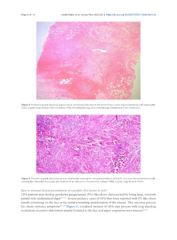
Figure 2. Photomicrograph depicting large zones of necrobiosis/necrosis in the dermis from a case of granulomatosis with polyangiitis
(H&E, original magnification 40×) (courtesy of the Dermatopathology Unit, Dermatology Department, Cairo University).
Figure 3. Photomicrograph depicting features of palisaded neutrophilic and granulomatous dermatitis in a case of granulomatosis with
polyangiitis: interstitial histiocytes and neutrophils as well as foci of necrobiotic collagen (H&E, original magnification 100×).
Rare or unusual clinical presentations of vasculitic skin lesions in AAV
GPA patients may develop pyoderma gangrenosum (PG)-like ulcers characterized by being large, intensely
painful with undermined edges [8,21,22] . Several pediatric cases of GPA have been reported with PG-like ulcers
mainly presenting on the face as the initial presenting manifestation of the disease. This can even precede
the classic systemic symptoms [23-27] [Figure 9]. Localized variants of GPA may present with long standing
recalcitrant ulcerative skin lesions mainly localized to the face and upper respiratory tract mucosa [28,29] .