
Page 61 - Read Online
P. 61
Abdel-Halim et al. Vessel Plus 2022;6:8 https://dx.doi.org/10.20517/2574-1209.2021.40 Page 3 of 14
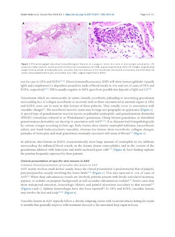
Figure 1. Photomicrograph depicting histopathological features of a purpuric lesion in a case of microscopic polyangiitis. (A)
Leukocytoclastic vasculitis involving small vessels in the subcutaneous fat (H&E, original magnification 40×). (B) A higher magnification
image showing details of leukocytoclastic vasculitis: fibrinoid necrosis of the vessel wall, neutrophils surrounding and infiltrating the
vessel, extravasated erythrocytes, and nuclear dust (H&E, original magnification 100×).
not the case in GPA and EGPA [10-12] . Direct immunofluorescence (DIF) will show immunoglobulin (usually
IgM) and complement C3 deposition around the walls of blood vessels in 70% and 50% of cases of GPA and
EGPA, respectively [8,13] . DIF is usually negative in MPA apart from possible few deposits of IgM and C3 [4,14] .
Granulomas which are extravascular in nature (usually necrobiotic palisading or necrotizing granulomas
surrounding foci of collagen necrobiosis or necrosis) such as those encountered in internal organs in GPA
and EGPA cases can be seen in skin lesions of these patients. They usually occur in association with
vasculitic changes . The necrobiotic/necrotic zones may be large and geographic in appearance [Figure 2].
[8]
A special type of granulomatous reaction known as palisaded neutrophilic and granulomatous dermatitis
(PNGD) (sometimes referred to as Winkelmann’s granuloma, Churg-Strauss granuloma, or interstitial
granulomatous dermatitis) can develop in association with AAV [15,16] . It is characterized histopathologically
by various changes according to their age. Early lesions show intense neutrophil infiltrates, karyorrhectic
debris, and frank leukocytoclastic vasculitis, whereas late lesions show necrobiotic collagen changes,
palisades of histiocytes, and small granulomas eventually associated with areas of fibrosis [Figure 3].
[17]
In addition, skin lesions in EGPA characteristically show large amount of eosinophils in the infiltrate
surrounding the inflamed blood vessels, in the dermis (tissue eosinophilia), and in the context of the
granulomas admixed with histiocytes and multi-nucleated giant cells [8,13] [Figure 4]. Such finding explains
the pruritus frequently expressed by these patients.
Clinical presentation of specific skin lesions in AAV
Common clinical presentations of vasculitic skin lesions in AAV
AAV mainly involves small dermal vessels, hence the clinical presentation is predominantly that of palpable
purpura/petechia usually involving the lower limbs [8,18] [Figure 5]. This was reported in 15% of cases of
AAV . When deep subcutaneous vessels are involved, patients present with livedo reticularis/racemosa,
[2]
[19]
papules, or nodules on purpuric background, as well as tender subcutaneous nodules . Severe cases may
show widespread extension, hemorrhagic blisters, and painful ulcerations secondary to skin necrosis
[6,7]
[2]
[Figures 6 and 7]. Splinter hemorrhages have also been reported . In GPA and EGPA, vasculitic lesions
may involve the face and scalp [5,8,20] [Figure 8].
Vasculitic lesions in AAV typically follow a chronic relapsing course with recurrent attacks lasting for weeks
to months that generally improve with treatment directed to the associated deep organ in focus.