


Page 52 - Read Online
P. 52
Inoue et al. Vessel Plus 2021;5:11  I  http://dx.doi.org/10.20517/2574-1209.2020.99                                                Page 5 of 10


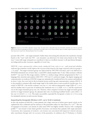
               Figure 3. Example of the RAPID mismatch output map. The pink lesion in the left shows the estimated ischemic core (18 mL), and the
               green lesion shows the penumbra (54 mL), which has a mismatch ratio of 3.0. These profiled cases are called target mismatch and have a
               high response to reperfusion therapy beyond the time window.

               patients with a PWI - core target mismatch. Rather than determining the indication for reperfusion therapy
               based on the “time” itself, the PWI - core mismatch can determine the brain status based on the “tissue
               time.” Cases with target mismatch are considered to have an excellent response to all reperfusion therapies,
               including endovascular treatment, regardless of onset time.


               DEFUSE 2 was a prospective cohort study conducted from 2008 to 2011 and examined whether
               endovascular treatment would improve the outcome determined by the target mismatch selection group in
                        [3]
               12 h onset . The target mismatch group was determined by DWI-PWI mismatch, and it was defined as a
               PWI/DWI mismatch ratio of more than 1.8 and DWI volume smaller than 70 mL. Fully automatic software
                     [30]
               RAPID  was used for this image analysis. RAPID is a medical image software programmed in the C ++
               language that calculates and analyzes MRI DWI / PWI and CT perfusion images. The digital imaging and
               communication in medicine (DICOM) images are automatically transferred from the scanner and analyzed
               in about 5 min, then sent back to the in-hospital picture archiving and communication systems (PACS). It
               is also possible to automatically email image maps to the stroke team or sent securely by mobile apps. As a
               result, in DEFUSE 2, the median ischemic core was 15.1 mL with an interquartile range (IQR) of 7.2-45.9,
               and the median time to puncture of endovascular treatment was 6.2 h (IQR: 4.9-8.1), and the reperfusion
               rate in the target mismatch group was 59%. However, when compared between the target mismatch group
               and the no target mismatch group, the adjusted odds ratio of achieving modified Rankin scale (mRS) 2 or
               less was 4.0 (95%CI: 1.3-12.2). This study successfully expanded the therapeutic window to 12 h by using
               the mismatch selection in endovascular therapy.


               Expanding the therapeutic Window in EVT, up to 16-24 h maximum
               In this sub-analysis of DEFUSE 2, some patients had a high variation of infarct grow speed, which can be
                                                                                                [31]
               explained by their collaterals and the existence of the penumbra within the time frame of 6-12 h . The rate
               of expansion of ischemic core lesions in early-onset DWI was highly variable. Patients with a malignant
               profile [32,33]  were found to have faster growth of ischemic core lesions than patients with target mismatch.
               Conversely, slow expansion of ischemic core lesions was associated with improved clinical outcome
               after reperfusion therapy with the recovery of the penumbra. In addition, the CT Perfusion to Predict

