
Page 40 - Read Online
P. 40
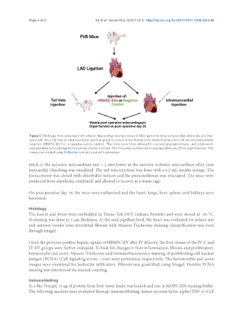
Page 4 of 9                      Xu et al. Vessel Plus 2023;7:33  https://dx.doi.org/10.20517/2574-1209.2023.98


                Figure 1. Wild-type mice underwent left anterior descending coronary artery (LAD) ligation to induce myocardial infarction, and then
                were split into a tail vein or intramyocardial injection group to receive either human bone mesenchymal stem cell-derived extracellular
                vesicles (HBMSC-EV) or a negative saline control. The mice were then allowed to recover postoperatively, and underwent
                postoperative echocardiogram to evaluate cardiac function. The mice were euthanized on postoperative day 28 for organ harvest. This
                image was created using BioRender.com and used with permission.

               stitch in the ischemic myocardium and 1-2 mm lower in the anterior ischemic myocardium after clear
               myocardial blanching was visualized. The tail vein injection was done with a 0.5 mL insulin syringe. The
               thoracotomy was closed with absorbable sutures and the pneumothorax was evacuated. The mice were
               awakened from anesthesia, extubated, and allowed to recover in a warm cage.


               On postoperative day 28, the mice were euthanized and the heart, lungs, liver, spleen, and kidneys were
               harvested.

               Histology
               The hearts and livers were embedded in Tissue-Tek OCT (Sakura Finetek) and were stored at -80 °C.
               Sectioning was done in 5 µm thickness. At the mid-papillary level, the heart was evaluated for infarct size
               and anterior border zone interstitial fibrosis with Masson-Trichrome staining. Quantification was done
               through ImageJ.

               Given the previous positive hepatic uptake of HBMSC-EV after IV delivery, the liver tissues of the IV-C and
               IV-EV groups were further evaluated. To look for changes in liver inflammation, fibrosis and proliferation,
               hematoxylin and eosin, Masson-Trichrome and immunofluorescence staining of proliferating cell nuclear
               antigen (PCNA) (Cell Signaling #2586, 1:400) were performed, respectively. The hematoxylin and eosin
               images were examined for leukocyte infiltration. Fibrosis was quantified using ImageJ. Positive PCNA
               staining was determined via manual counting.

               Immunoblotting
               In a Bis-Tris gel, 10 μg of protein from liver tissue lysate was loaded and run in MOPS-SDS running buffer.
               The following markers were evaluated through immunoblotting: tumor necrosis factor alpha (TNF-α) (Cell