
Page 43 - Read Online
P. 43
Xu et al. Vessel Plus 2023;7:33 https://dx.doi.org/10.20517/2574-1209.2023.98 Page 7 of 9
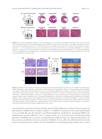
Figure 3. There were no significant changes in myocardial infarct size or border zone interstitial fibrosis after either IM or IV human
bone mesenchymal stem cell-derived extracellular vesicle (HBMSC-EV) injection. (A) The infarct percentages of all four groups were
not found to be significantly different (P = 0.05620). Representative images of the infarct size in each of the four groups are shown.
Scale bar = 1 mm. (B) Anterior border zone interstitial fibrosis was not found to be significantly different (P = 0.6333).
Scale bar = 100 µm; IM-C, IM control injection group; IM-EV, IM extracellular vesicle injection group; IV-C, IV control injection group;
IV-EV, IV extracellular vesicle injection group. The Shapiro-Wilk test was used to determine that the data were parametric. Then, the
one-way ANOVA test was performed.
Figure 4. Intravenous (IV) injection of human bone mesenchymal stem cell-derived extracellular vesicles (HBMSC-EV) did not alter
hepatic inflammatory status, fibrosis, or proliferation. (A) Hematoxylin and eosin staining, Masson-Trichrome staining, and proliferating
cell nuclear antigen (PCNA) immunofluorescence did not demonstrate differences in leukocyte infiltration, fibrosis, and cellular
proliferation, respectively. Scale bar = 100 µm. (B) Quantification of % fibrosis on Masson-Trichrome staining was not significantly
different (P = 0.8167) between the IV control (IV-C) and IV HBMSC-EV (IV-EV) injection groups. Statistical analysis was performed
using the Shapiro-Wilk and unpaired t-test. (C) Immunoblotting showed no differences in major regulators of inflammatory and fibrosis
pathways, such as tumor necrosis factor-alpha (TNF-α), transforming growth factor-β (TGF-β), and SMAD 2/3. Cell proliferation
regulator mammalian target of rapamycin (mTOR) expression was increased in the IV-EV group, but this finding is difficult to interpret
given the complexity of mTOR regulation and the negative PCNA findings. Oxidative protein expression changes are difficult to
interpret as well, given the lack of hepatic oxidative injury in this model.
even among EVs isolated from similar cell types, given the likely differences in donors. However, given the
9
large number of HBMSC that we needed to isolate 2 × 10 HBMSC-EV particles for this small animal model,
we do not think that our dose should be considered insufficient. Furthermore, optimizing HBMSC-EV
administration timing is difficult to do in a murine model, as re-do thoracotomy and intramyocardial
injection is prohibitive due to operative scarring from the initial procedure. In a large animal model, re-do
thoracotomy is possible but is far more expensive. If HBMSC-EV were able to be effectively delivered to the
heart non-invasively, there would be far more flexibility in testing dosing timing. Perhaps also a cardiac