


Page 37 - Read Online
P. 37
Page 6 of 9                     Buia et al. Vessel Plus 2022;6:48  https://dx.doi.org/10.20517/2574-1209.2022.04


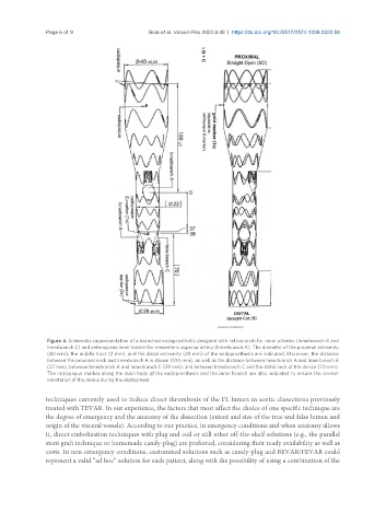
                Figure 4. Schematic rappresentation of a branched endoprosthetis designed with retrobranch for renal arteries (Innerbranch B and
                Innerbranch C) and anterograde inner branch for mesenteric superior artery (Innerbranch A). The diameter of the proximal extremity
                (40 mm), the middle tract (2 mm), and the distal extremity (28 mm) of the endoprosthesis are indicated. Moreover, the distance
                between the proximal neck and Innerbranch A is shown (100 mm), as well as the distance between Innerbranch A and Innerbranch B
                (37 mm), between Innerbranch A and Innerbranch C (39 mm), and between Innerbranch C and the distal neck of the device (70 mm).
                The radiopaque marker along the main body of the endoprosthesis and the inner branch are also indicated to ensure the correct
                orientation of the device during the deployment.

               techniques currently used to induce direct thrombosis of the FL lumen in aortic dissections previously
               treated with TEVAR. In our experience, the factors that most affect the choice of one specific technique are
               the degree of emergency and the anatomy of the dissection (extent and size of the true and false lumen and
               origin of the visceral vessels). According to our practice, in emergency conditions and when anatomy allows
               it, direct embolization techniques with plug and coil or still other off-the-shelf solutions (e.g., the parallel
               stent graft technique or homemade candy-plug) are preferred, considering their ready availability as well as
               costs. In non-emergency conditions, customized solutions such as candy-plug and BEVAR/FEVAR could
               represent a valid “ad hoc” solution for each patient, along with the possibility of using a combination of the

