
Page 96 - Read Online
P. 96
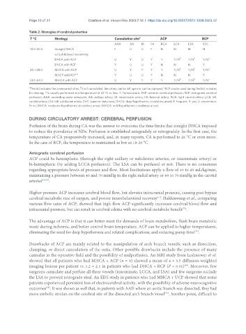
Page 10 of 21                Calafiore et al. Vessel Plus 2023;7:18  https://dx.doi.org/10.20517/2574-1209.2023.42

               Table 2. Strategies of cerebral protection
                                                                    a
                T °C       Strategy                                                                Cannulation site                                               ACP                         RCP
                                                        AAA     AA   IA   FA   RCA   LCA    LSA    SVC
                14.0-20.0  Straight DHCA                F       U    U    F    N     N      N      N
                           w/out delayed rewarming
                                                                                        b      b
                           DHCA with ACP                U       Y    U    Y    Y     Y/N    Y/N    Y/N*
                           DHCA with RCP                Y       U    U    Y    N     N      N      Y
                20.1-28.0  MHCA with ACP                U       Y    Y    Y    Y     Y/N b  Y/N b  Y/N*
                           MHCP with RCP**              Y       U    U    Y    N     N      N      Y
                28.1-34.0  MiHCA with ACP               U       Y    Y    Y    Y     Y/N b  Y/N b  Y/N*
               a                        b
                The list includes the commonest sites;  if not cannulated: the artery can be left open or can be clamped; *RCP can be used during the last minutes
               for dearing; **is usually performed at a temperature of 22 °C or less. T: Temperature; ACP: anterior cerebral perfusion; RCP: retrograde cerebral
               perfusion; AAA: ascending aorta aneurysm; AA: axillary artery; IA: innominate artery; FA: femoral artery; RCA: right carotid artery; LCA: left
               carotid artery; LSA: left subclavian artery; SVC: superior vena cava; DHCA: deep hypothermic circulatory arrest; F: frequent; Y: yes; U: uncommon;
               N: no; MHCA: moderate hypothermic circulatory arrest; MiHCA: mild hypothermic circulatory arrest.

               DURING CIRCULATORY ARREST: CEREBRAL PERFUSION
               Perfusion of the brain during CA was the answer to overcome the time limits that straight DHCA imposed
               to reduce the prevalence of NDs. Perfusion is established antegradely or retrogradely. In the first case, the
               temperature of CA progressively increased, and, in many reports, CA is performed to 26 °C or even more.
               In the case of RCP, the temperature is maintained as low as 18-20 °C.


               Antegrade cerebral perfusion
               ACP could be hemispheric (through the right axillary or subclavian arteries, or innominate artery) or
               bi-hemispheric (by adding LCCA perfusion). The LSA can be perfused or not. There is no consensus
               regarding appropriate levels of pressure and flow. Most Institutions apply a flow of 10 to 20 mL/kg/min,
               maintaining a pressure between 40 and 70 mmHg in the right radial artery or 60 to 70 mmHg in the carotid
               arteries [69,70] .

               Higher pressure ACP increases cerebral blood flow, but elevates intracranial pressure, causing post bypass
               cerebral metabolic rate of oxygen, and poorer neurobehavioral recovery . Haldenwang et al., comparing
                                                                             [71]
               various flow rates of ACP, showed that high-flow ACP significantly increases cerebral blood flow and
               intracranial pressure, but can result in cerebral edema with no cerebral metabolic benefit .
                                                                                         [72]

               The advantage of ACP is that it can better meet the demands of brain metabolism, flush brain metabolic
               waste during ischemia, and better control brain temperature. ACP can be applied to higher temperatures,
                                                                                               [73]
               eliminating the need for deep hypothermia and related complications, and reducing pump time .

               Drawbacks of ACP are mainly related to the manipulation of arch branch vessels, such as dissection,
               clamping, or direct cannulation of the ostia. Other possible drawbacks include the presence of many
               cannulas in the operative field and the possibility of malperfusion. An MRI study from Leshnower et al.
               showed that all patients who had MHCA + ACP (n = 9) showed a mean of 4 ± 3.5 diffusion-weighted
               imaging lesions per patient vs. 1.2 ± 2.1 in patients who had DHCA + RCP (P = 0.01) . Moreover, few
                                                                                          [36]
               surgeons cannulate and perfuse all three vessels (innominate, LCCA, and LSA) and few surgeons occlude
               the LSA to prevent retrograde steal. An EEG study in patients who had MHCA + UCP showed that some
               patients experienced persistent loss of electrocerebral activity, with the possibility of adverse neurocognitive
                       [74]
               outcomes . It was shown as well that, in patients with AAD where an aortic branch was dissected, they had
               more embolic strokes on the cerebral site of the dissected arch branch vessel . Another point, difficult to
                                                                                 [75]