


Page 128 - Read Online
P. 128
Page 6 of 19                                                         Chen et al. Soft Sci. 2026, 6, 48


               port. Scattering Boundary Condition was added to the air boundary. Variation of the heating distance was
               simulated by adjusting the distance between the port and the CNT-coated balloon, and the heating power
               could also be simulated by setting the port power. In the Heat Transfer in Solids (ht) section, set the initial
               system temperature to 293.15 K. In the Mesh section, the grids were created. Finally, the microwave
               frequency in the Frequency Domain was set to 2,450 MHz, and the heating time in Transient was set to
               2 min in this study. Then the model was submitted to calculate and perform post-processing on the results.


               Magnetic deflection simulation of the MBF
               Simulations of the magnetic deflection angles θ of the MBF in uniform magnetic fields, with varying field
               strengths B and angles α, were performed by implementing a user-defined element (UEL) in
               Abaqus/Standard 2023. To optimize computational efficiency without compromising modeling accuracy, the
               external balloon (elastic modulus ~100 kPa) was excluded from the model, as its mechanical resistance
               during deflection is negligible compared to the nitinol core (40 GPa). For the retained structures, the nitinol
               core was modeled as an elastic material with a Young’s modulus of 40 GPa and a Poisson’s ratio of 0.3, and
               was meshed by 856 C3D8R elements. The magnetic composite was modeled using a hyperelastic material
               with a shear modulus of 850 kPa and meshed by 15,024 UEL elements. Its bulk modulus was set to 1,000
               times the shear modulus to approximate the material’s incompressibility, and its magnetization was defined
               as 128 kA·m . Finally, the strength (field strength B) and direction (angle α) of the magnetic fields, along with
                         -1
               the magnetization, were incorporated as additional input parameters.


               Biocompatibility test of the MBF
               The experimental design for this study aimed to evaluate the biological response of L-929 cells to the MBF.
               The test samples (magnetic tip and phase-change balloon) were subjected to an extract preparation process
               under sterile conditions. Specifically, the samples were immersed in 75.6 mL of MEM medium containing
               10% fetal bovine serum at a ratio of 3 cm  of sample surface area to 1 mL of extractant. The immersion was
                                                  2
               conducted in a sterile, inert container at 37 °C for 48 h with oscillation. After immersion, the extract was
               checked for any changes and stored at 20-30 °C, ready for use within 24 h without further filtration,
               centrifugation, dilution, or pH adjustment. Simultaneously, blank, negative (high-density polyethylene), and
               positive (ZDEC) controls were prepared under the same conditions. The L-929 cells, sourced from the
               American Type Culture Collection (CCL1, NCTC clone 929), were cultured in MEM medium supplemented
               with 10% fetal bovine serum and antibiotics (penicillin and streptomycin) at 37 °C in a 5% CO  incubator.
                                                                                                2
               Following cell growth to confluence, the original medium was aspirated, and 100 μL of different
               concentrations of the test sample extract (100%, 75%, 50%, 25%), blank control, positive control (100%), and
               negative control (100%) were added to the cells. The cells were then incubated for an additional 24 h at 37 °C
               in a 5% CO  atmosphere.
                        2

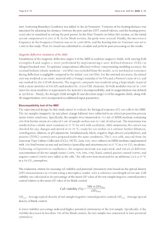
               The evaluation criteria for assessing cell viability and potential cytotoxicity were based on the optical density
               (OD) measurements at 570 nm using a microplate reader, with a reference wavelength of 650 nm. Cell
               viability was calculated as the percentage of the mean OD value of the test sample/negative control/positive
               control relative to the mean OD value of the blank control.


                                            Cell viability (%) =


               OD 570e  - Average optical density of test sample/negative control/positive control; OD 570b  - Average optical
               density of blank control.

               A lower viability percentage indicated higher potential cytotoxicity of the test sample. Specifically, if the
               viability decreased to less than 70% of the blank control, the test sample was considered to have potential
               cytotoxicity.

