
Page 89 - Read Online
P. 89
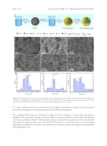
Page 4 of 16                            Sun et al. Soft Sci. 2025, 5, 35  https://dx.doi.org/10.20517/ss.2025.21


                Figure 1. (A) Schematic synthesis of annealed FeCoNi@ZnIn S  microspheres with core-shell structure; SEM images of (B) ZnIn S , (C)
                                                        2 4
                                                                                                       4
                                                                                                     2
                FeCoNi, (D) FeCoNi@ZnIn S , (E) FCNZ-500, (F) FCNZ-600 and (G) FCNZ-700 microspheres; the particle size distribution of (H)
                                   4
                                  2
                FCNZ-500, (I) FCNZ-600 and (J) FCNZ-700 microspheres. SEM: Scanning electron microscope.
                 3+
                        2-
               In , and S  and then heated by an oil bath for 2 h. The ZnIn S  nanosheets as a dielectric shell were grown
                                                                   2 4
               on FeCoNi microspheres and subsequently annealed at different temperatures.
               The resulting SEM images are displayed in Figure 1B-G. Pure ZnIn S  is a flower-like microsphere
                                                                             2 4
               assembled from numerous irregular nanosheets. Different diameters and sizes of flower-like microspheres
               can be seen inside the cluster . Subsequently, ZnIn S  nanosheets were grown on the rough FeCoNi
                                          [35]
                                                              2 4
               microspheres, which indicates the core-shell FCNZ structure. At the same time, as the annealing
               temperature increased, the microspheres gradually became smoother, and the average particle size gradually
               increased [Figure 1H-J].