
Page 830 - Read Online
P. 830
Page 6 of 14 Mirastschijski et al. Plast Aesthet Res 2020;7:70 I http://dx.doi.org/10.20517/2347-9264.2020.147
M
L
K N
O
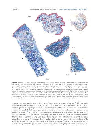
Figure 2. Microstructure of the skin from different body areas. A: lower left arm, B: penis, C: small labia. Note the dense dermal
structure with multiple vessels in (A) and lose collagen bundles in (B) and (C) with high similarity of penile and labial skin. D: panel HE
staining, E and F: Elastica van Gieson staining. Arrow upper panel depicting epidermis, pD papillary dermis, rD reticular dermis. Note
abundance of collagen fibres in arm skin (red staining, black arrow; panel E and F) and abundant elastic fibres in penile and labial skin
(black staining, white arrows). Scale bar in A for upper two panels 500 µm, lower panel 40× magnification. G-O: Masson Trichrome
staining for visualization of collagen fibres (blue staining) in normal and scar tissues of different origin. G, H, I, N normal skin, K, L, M, O
scar tissue; G and K breast, H and L scrotum, I and M penis, N and O abdomen. Note abundant collagen bundles in normal breast and
abdominal skin (G and N) and densely packed collagen bundles in scar tissue of breast and abdomen (K and O, respectively). Thickened
dermal tissue in breast scar (K). Loose connective tissue with scrotal (H, L) and penile (I, M) normal and scar tissue without densely
packed collagen bundles as seen in non-genital scar tissue. Scale bars next to each specimen indicating magnification
[11]
example, oestrogens accelerate wound closure, whereas testosterone delays healing . Skin is a major
source of extra-glandular sex steroid hormones. The intracellular enzyme aromatase converts the sex
hormone precursor dehydroepiandrosterone downstream into estrone or via testosterone into the more
potent 17β-oestradiol. Both oestrogens act via the oestrogen receptors and stimulate keratinocyte and
[10]
fibroblast migration . In genital fibroblasts, aromatase expression is androgen dependent and oestrogens
stimulate fibroblast contractility without increasing alpha-smooth muscle actin expression or myofibroblast
differentiation . Upon wounding, aromatase activity increases 400-fold in keratinocytes with increased
[12]
intracellular oestrogens. Oestrogens reduce the cellular inflammatory response via downregulation of the
pro-inflammatory cytokine macrophage migration inhibitory factor , by reduced toll-like-receptor-4
[13]
mediated mitogen activated protein kinase activation, by reduced macrophage infiltration into wounds and