
Page 828 - Read Online
P. 828
Page 4 of 14 Mirastschijski et al. Plast Aesthet Res 2020;7:70 I http://dx.doi.org/10.20517/2347-9264.2020.147
Table 1. Skin structure of homologous male and female outer genitalia
Male Female Histological microstructure
Glans Of penis* Of clitoris Multilayered, non-keratinizing epidermis, dermal tissue with abundant nerve
endings
Foreskin Of penis Of clitoris Outer part: epidermis with cornified layer
Inner part: non-keratinizing epidermis; mucous epithelium; no fat tissue
Frenulum Frenulum penis Frenula clitoridis (paired) Non-keratinizing, mucous epithelium, no subcutaneous fat tissue
Penile shaft skin Small labia Penis: epidermis with cornified layer, highly flexible attachment to underlying
tissue via Dartos fascia (Fascia penis superficialis)
Labia: outer surface with thin cornified layer; inner surface: no cornified layer
Both: no hair; no fat tissue; many elastic fibers
Scrotum Big labia Hair bearing epidermis (labia: only outer surface), epidermal cornified layer
Labia: subcutaneous fat layer and smooth muscle cells
Scrotum: no (or very little) fat, but contractile Tunica Dartos with smooth
muscle cells and myofibroblasts; in obese patients: fat tissue
*After circumcision, the epithelium changes into a keratinizing epidermis of the glans penis
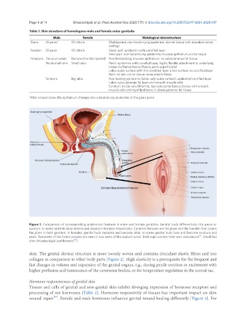
Figure 1. Comparison of corresponding anatomical features in male and female genitalia. Genital buds differentiate into penis or
scrotum in males and into labia minora and majora in females respectively. Common features are the glans and the foreskin that covers
the glans in both genders. In females, genital buds separate and become labia, in males genital buds fuse and become scrotum and
[8]
penis. Remnants of the fusion process are seen in two parts of the septum scroti. Both septi contain their own vasculature . (modified
[9]
after Mirastschijski and Remmel )
skin. The genital dermal structure is more loosely woven and contains abundant elastic fibres and less
collagen in comparison to other body parts [Figure 2]. High elasticity is a prerequisite for the frequent and
fast changes in volume and expansion of the genital organs, e.g., during penile erection or excitement with
higher perfusion and tumescence of the cavernous bodies, or for temperature regulation in the scrotal sac.
Hormone responsiveness of genital skin
Tissues and cells of genital and non-genital skin exhibit diverging expression of hormone receptors and
processing of sex hormones [Table 2]. Hormone responsivity of tissues has important impact on skin
[10]
wound repair . Female and male hormones influence genital wound healing differently [Figure 3]. For