
Page 804 - Read Online
P. 804
Page 8 of 12 Bair et al. Plast Aesthet Res 2020;7:68 I http://dx.doi.org/10.20517/2347-9264.2020.74
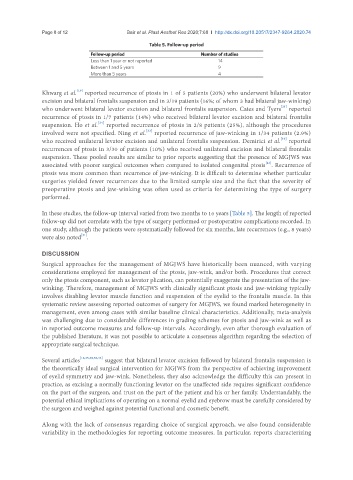
Table 5. Follow-up period
Follow-up period Number of studies
Less than 1 year or not reported 14
Between 1 and 5 years 9
More than 5 years 4
[19]
Khwarg et al. reported recurrence of ptosis in 1 of 5 patients (20%) who underwent bilateral levator
excision and bilateral frontalis suspension and in 3/19 patients (16%; of whom 3 had bilateral jaw-winking)
[21]
who underwent bilateral levator excision and bilateral frontalis suspension. Cates and Tyers reported
recurrence of ptosis in 1/7 patients (14%) who received bilateral levator excision and bilateral frontalis
[24]
suspension. Ho et al. reported recurrence of ptosis in 2/8 patients (25%), although the procedures
[32]
involved were not specified. Ning et al. reported recurrence of jaw-winking in 1/34 patients (2.9%)
[16]
who received unilateral levator excision and unilateral frontalis suspension. Demirici et al. reported
recurrences of ptosis in 3/30 of patients (10%) who received unilateral excision and bilateral frontalis
suspension. These pooled results are similar to prior reports suggesting that the presence of MGJWS was
associated with poorer surgical outcomes when compared to isolated congenital ptosis . Recurrence of
[23]
ptosis was more common than recurrence of jaw-winking. It is difficult to determine whether particular
surgeries yielded fewer recurrences due to the limited sample size and the fact that the severity of
preoperative ptosis and jaw-winking was often used as criteria for determining the type of surgery
performed.
In these studies, the follow-up interval varied from two months to 16 years [Table 5]. The length of reported
follow-up did not correlate with the type of surgery performed or postoperative complications recorded. In
one study, although the patients were systematically followed for six months, late recurrences (e.g., 8 years)
[21]
were also noted .
DISCUSSION
Surgical approaches for the management of MGJWS have historically been nuanced, with varying
considerations employed for management of the ptosis, jaw-wink, and/or both. Procedures that correct
only the ptosis component, such as levator plication, can potentially exaggerate the presentation of the jaw-
winking. Therefore, management of MGJWS with clinically significant ptosis and jaw-winking typically
involves disabling levator muscle function and suspension of the eyelid to the frontalis muscle. In this
systematic review assessing reported outcomes of surgery for MGJWS, we found marked heterogeneity in
management, even among cases with similar baseline clinical characteristics. Additionally, meta-analysis
was challenging due to considerable differences in grading schemes for ptosis and jaw-wink as well as
in reported outcome measures and follow-up intervals. Accordingly, even after thorough evaluation of
the published literature, it was not possible to articulate a consensus algorithm regarding the selection of
appropriate surgical technique.
Several articles [16,25,28,30,33] suggest that bilateral levator excision followed by bilateral frontalis suspension is
the theoretically ideal surgical intervention for MGJWS from the perspective of achieving improvement
of eyelid symmetry and jaw-wink. Nonetheless, they also acknowledge the difficulty this can present in
practice, as excising a normally functioning levator on the unaffected side requires significant confidence
on the part of the surgeon, and trust on the part of the patient and his or her family. Understandably, the
potential ethical implications of operating on a normal eyelid and eyebrow must be carefully considered by
the surgeon and weighed against potential functional and cosmetic benefit.
Along with the lack of consensus regarding choice of surgical approach, we also found considerable
variability in the methodologies for reporting outcome measures. In particular, reports characterizing