
Page 718 - Read Online
P. 718
Page 6 of 12 Meltzer et al. Plast Aesthet Res 2020;7:61 I http://dx.doi.org/10.20517/2347-9264.2020.122
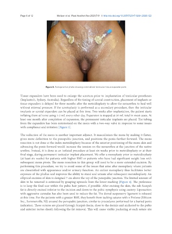
Figure 3. Postoperative photo showing externalized testicular tissue expander ports
Tissue expanders have been used to enlarge the scrotum prior to implantation of testicular prostheses
(Implantech, Sydney, Australia). Regardless of the timing of scrotal construction, placement of implants or
tissue expanders is delayed for three months after the metoidioplasty to allow the neourethra to heal well
without external pressure. If the scrotoplasty is performed as a secondary procedure, then the testicular
implants or scrotal expanders can be placed at this time. Two weeks after implantation, the patient starts
inflating them at home using 1-2 mL every other day. Expansion is stopped at 50 mL total in most cases. At
least one month after completion of expansion, the permanent testicular implants are placed. The tubing
from the expanders has been exteriorized on the mons with a two-way valve in response to some issues
with compliance and irritation [Figure 3].
The reduction of the mons is another important adjunct. It masculinizes the mons by making it flatter,
gives more definition to the penopubic junction, and positions the penis further forward. The mons
resection is not done at the index metoidioplasty because of the anterior positioning of the mons skin and
advancing the penis forward would increase the tension on the neourethra at the junction of the native
urethra. Instead, it is done as an isolated procedure at least six weeks prior to metoidioplasty or at their
final stage, during permanent testicular implant placement. We offer a monsplasty prior to metoidioplasty
(at least six weeks) for patients with higher BMI or patients who have had significant weight loss with
subsequent mons ptosis. The mons resection in this group will need to be a more extended excision. By
performing this procedure, we try to avoid some of the issues that arise after monsplasty where patients
are dissatisfied with appearance and/or urinary function. An earlier monsplasty thus facilitates better
exposure of the phallus and improves the ability to stand and urinate after subsequent metoidioplasty. An
elliptical excision of skin is designed 7 cm above the top of the penopubic junction. The limited amount of
skin to be removed is estimated by grasping upwards from the lower marking [Figure 4]. The preference
is to keep the final scar within the pubic hair pattern, if possible. After excising the skin, the sub-Scarpa’s
fat is directly excised inferior to the incision and down to the pubic symphysis using cautery. Liposuction
with aggressive cannulas has also been used to reduce the fat. The dorsal suspensory ligament is released
at this time. For the patients with a greater BMI, they benefit from tacking sutures with 0 Prolene (Ethicon
Inc., Sommerville, NJ) around the penopubic junction, similar to procedures performed for a buried penis
indication. These sutures are placed through Scarpa’s fascia, close to the dermis and anchored to the pubis
and anterior rectus sheath following the fat removal. This will cause visible puckering at each suture site