
Page 494 - Read Online
P. 494
Zein et al. Plast Aesthet Res 2020;7:44 I http://dx.doi.org/10.20517/2347-9264.2020.133 Page 3 of 10
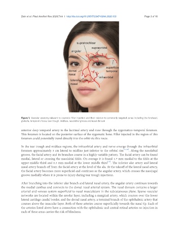
Figure 1. Vascular anatomy relevant to cosmetic filler injection and their relation to commonly targeted zones including the forehead,
glabella, temporalis fossa, tear trough, midface, nasolabial groove and nasal dorsum
anterior deep temporal artery to the lacrimal artery and runs through the zygomatico-temporal foramen.
This foramen is located on the posterior surface of the zygomatic bone. Filler injected in the region of this
foramen could potentially travel directly into the orbit via this route.
In the tear trough and midface regions, the infraorbital artery and nerve emerge through the infraorbital
foramen approximately 3 cm lateral to midline just inferior to the orbital rim [11,12] . Along the nasolabial
groove, the facial artery and its branches course in a highly variable pattern. The facial artery can be found
medial, lateral or crossing the nasolabial folds. On average it is found 1.7 mm medial to the folds at the
[13]
upper middle third and 0.3 mm medial at the lower middle third . The inferior alar artery and lateral
nasal artery branch off from the facial artery at the level of the ala. At the takeoff of the lateral nasal artery,
the facial artery becomes more superficial and continues as the angular artery, which crosses the nasojugal
groove medially where it is prone to injury during tear trough injections.
After branching into the inferior alar branch and lateral nasal artery, the angular artery continues towards
the medial canthus and connects to the dorsal nasal arterial system. The nasal dorsum contains a larger
arterial and venous system superficial to nasal musculature in the subcutaneous plane. Sparse vascular
networks are located within the areolar layer, including a marginal artery, which courses over the lower
lateral cartilage caudal border, and the dorsal nasal artery, a terminal branch of the ophthalmic artery that
courses above the muscular layer. Both of these arteries course superficially towards the nasal tip. Each of
the arteries listed above have a connection with the ophthalmic and central retinal arteries so injection in
each of these areas carries the risk of blindness.