
Page 469 - Read Online
P. 469
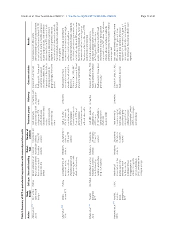
Citterio et al. Plast Aesthet Res 2020;7:41  I  http://dx.doi.org/10.20517/2347-9264.2020.29                                     Page 13 of 20


                        Sites treated with PDL grafts   in PDL grafts  Both groups showed a significant   without statistically significant   cells sheets were reported The test protocol resulted in a   of CAL gains, PPD reductions   were reported   PD reduction, CAL gain and bone   reported
                     Results  demonstrated significant improvement  in vertical and horizontal defect fill, PD,  and CAL at 3 and 6 months compared  to pre-surgical values. The difference  determined for the PD values of both  groups at a statistically significant  degree in favor of grafted sites was  maintained at all observation periods.  No foreign body reaction was observed   increase in the alveolar bone height,  differences between groups. Regarding  the clinical p


                     Outcome variables  Clinical: PI, GI, PD, GR,   Radiographic: linear and   volumetric evaluation  Volumetric defect fill by   impression of the defects  Histologic analysis by   gingival biopsy from one   patient  Radiographic (main   outcome): Increase in  alveolar bone height (rx   bone fill)  Clinical: CAL, PPD, REC Safety assessment: blood   and urine examination  Clinical: PI, BPI, CAL, PPD,  relative gingival marginal   level  Radiographic: linea


                     Follow-up  6 months, with   CAL a surgical re-  entry  12 months  6 months  12 months  CAL


                     Treatment groups  Test: coronally   positioned flap with   autogenous PDL   grafts that were   obtained from third   molars  Control: coronally   positioned flap   alone  Test: GTR and   PDLSC sheets in   combination with      demineralized   bovine bone matrix  Control: GTR and   demineralized   bovine bone matrix  without stem cells   Test: ODF applying   allogeneic UC-  MSCs on a   β-TCP scaffold in   combination rh-  PDGF-BB  Control: OFD  Test: m


                    Number of   patients  10 patients 20   10test/10   control  30 patients 41   defects:  20 test/21   control  14 patients  24 defects:  12 test/ 12   control  29 patients  29 defects:  15 test/14   control
                 Table 3. Summary of RCT on periodontal regeneration with mesenchymal stem cells


                    Defect   type  Class II   mandibular  defects:  furcation   defects  Intrabony   defects  Intrabony   defects  Intrabony   defects


                     Stem cells handling  Direct application of   PDL tissue collected   from an extracted   molar, into the   defect  Collection from an   extracted molar,   isolation, culture,   characterization and   engineering into cells   sheets in laboratory  Collection from the   hospital in a sterile   tube Followed by   isolation and culture   on β-TCP scaffold  Mechanical   dissociation of the   dental pulp of an   extracted tooth   by the use of a   biological


                     Cell type  PDLSC       PDLSC             UC-MSC           DPSC


                    Study   design  RCT with a   split mouth   design  single-  center RCT  Parallel   designed   RCT  double-  blind,   prospective   RCT


                        Akbay et al. [112]    Chen et al. [120]    Dhote et al. [123]    Ferrarotti et al. [122]   Parallel,

                     Author  2005             2016              2015             2018