
Page 299 - Read Online
P. 299
Evans et al. Plast Aesthet Res 2020;7:28 I http://dx.doi.org/10.20517/2347-9264.2019.53 Page 3 of 10
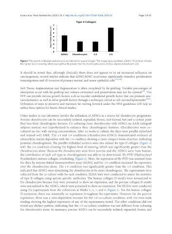
Figure 1. The paraffin embedded sections were also stained for type II collagen. The images were quantified, and the 1:9 condition showed
the highest level of staining, which was significantly greater than the chondrocytes alone. ADSCs: adipose derived stem cells
It should be noted that, although clinically there does not appear to be an increased influence on
carcinogenesis, several studies indicate that ADSC/ADSC secretomes significantly stimulate proliferation
transmigration and 3D invasion of primary normal and tumor epithelial cells [118-120] .
Soft Tissue Augmentation and Regeneration is often completed by fat grafting. Variable percentages of
[25]
absorption occur with fat grafting and volume estimation and preservation may not be optimal . The
SVF can provide various growth factors such as vascular endothelial growth factor that can promote neo-
vascularization as well as other growth factors through a technique coined as cell-assisted lipotransfer [62,63] .
Utilization of ways to preserve and maintain fat moving forward under the FDA guidelines will help us
utilize these options for future clinical studies.
Other studies in our laboratory involve the utilization of ADSCs as a source for chondrocyte progenitors.
Porcine chondrocytes can be successfully isolated, expanded, frozen, and thawed, but past a certain point
they lose their chondrogenic features. Co-culturing these chondrocytes with ADSCs on AAM (allograft
adipose matrix) was hypothesized to enhance their chondrogenic features. Chondrocytes were co-
cultured on disc with varying concentrations. After 10 weeks in culture, the discs were paraffin imbedded
and stained with H&E. The 1:5 and 1:9 conditions (chondrocytes:ADSCs) demonstrated evidence of
extracellular matrix deposition with the 1:9 condition showing a more compact tissue structure, indicating
potential chondrogenesis. The paraffin imbedded sections were also stained for type II collagen [Figure 1]
with the 1:9 condition showing the highest level of staining, which was significantly greater than the
chondrocytes alone. Because the chondrocytes were from porcine and the ADSCs were from human,
the contribution of each cell type to chondrogenesis was able to be determined. Hy-PYD (Hydroxylysyl
Pyridinoline) assesses collagen crosslinking [Figure 2]. Here, the expression of Hy-PYD was assessed from
the discs by enzyme-linked immunosorbent assay (ELISA) and the 1:9 condition increased the expression
over the chondrocytes alone. The 1:9 condition was significantly greater than the 1:5 condition. This
indicated that ADSCs were stimulating the chondrocytes to be more chondrogenic. The supernatants were
collected from the co-culture wells for each condition. ELISA tests were conducted to assess the secretion
of type II collagen using species specific antibodies. The human collagen II results were normalized to
the chondrocytes because they were presumed to show no expression, and the porcine collagen II results
were normalized to the ADSCs, which were presumed to show no expression. The ELISAs were conducted
using the supernatants from the collections at Weeks 3, 5, 7, and 9 [Figure 3]. For the human collagen
II expression, there was essentially no expression throughout the experiment. However, for the porcine
expression, there was a time-dependent increase for the 1:9 co-culture condition, with the nine-week
reading showing the highest expression of any of the supernatants tested. The other conditions did not
reveal any distinct pattern, indicating that the 1:5 co-culture condition was not different from culturing
the chondrocytes alone. In summary, porcine ADSCs can be successfully isolated, expanded, frozen, and