
Page 294 - Read Online
P. 294
Page 4 of 6 Kumar. Plast Aesthet Res 2020;7:27 I http://dx.doi.org/10.20517/2347-9264.2020.07
A B
C D
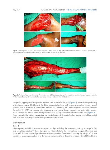
Figure 1. Photographs of case 1 showing: A: exposed Gortex vascular implant; B: limited access dressing covering the wound; C:
granulation covering implant after 20 days; D: 3 months after resurfacing by skin graft
A
B C
Figure 2. Photographs of case 2 showing: A: extensive wound following debridement for necrotizing fascitis; B: wound preparation under
limited access dressing; C: final result after resurfacing without development of distal edema
the patella, upper part of the patellar ligament, and subpatellar fat pad [Figure 3]. After thorough cleaning
and minimal wound debridement, the defect was partially closed with sutures as complete closure was not
possible, due to insertion of a tube drain and tubular LAD along with application of a posterior plaster of
Paris slab. The LAD was changed after 10 days and final wound closure was achieved under slight tension.
After 20 days, the patient was discharged with linear healing of the wound at the suture site [Figure 3C].
After 1 month, the patient was advised for physiotherapy. At 3 months’ follow-up, the wound had healed
well with mild hypertrophy and full range of motion of the knee.
DISCUSSION
Case 1
Major options available in this case were pedicled flaps including the latissimus dorsi flap, subscapular flap
[8]
and lateral thoracic flap . These flaps provide relative bulk to the recipient site compared to a SSG and
come with donor site related problems such as compromised function and scarring. By using LAD, it was
possible to achieve granulation over the Gortex implant and later, definitive coverage with a SSG in 28 days