Page 293 - Read Online
P. 293
Kumar. Plast Aesthet Res 2020;7:27  I  http://dx.doi.org/10.20517/2347-9264.2020.07                                                   Page 3 of 6

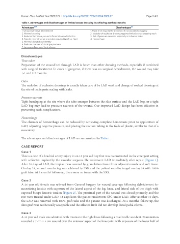
               Table 1. Advantages and disadvantages of limited access dressing in achieving aesthetic results
               Advantages [3-7]                                              Disadvantages [3]
               1. Ultraconservative debridement              1. More time required for treatment if not assisted by surgery
               2. Minimal scarring                           2. Malodor of occlusive dressing requires limited access dressing wash
               3. Reduces flap failure, wound inflammation and infection   3. Risk of pressure necrosis, especially in ischemic limbs
               4. Simpler reconstructive procedure required (graft vs. flap)   4. Hemorrhage
               5. Minimal donor site deformity
               6. Reduces chances of distal lymphedema
               7. Increases chances of limb salvage

               Disadvantages
               Time taken
               Preparation of the wound bed through LAD is faster than other dressing methods, especially if combined
               with surgical treatment. In cases of gangrene, if there was no surgical debridement, the wound may take
               1-1 and 1/2 months.

               Odor
               The malodor of occlusive dressings is usually taken care of by LAD wash and change of soaked dressings at
               the site of inadequate sealing with leaks.

               Pressure necrosis
               Tight bandaging at the site where the tube emerges between the skin surface and the LAD bag, or a tight
               LAD bag may lead to pressure necrosis of the wound. Our improved LAD design has been effective in
               preventing such complications.


               Hemorrhage
               The chances of hemorrhage can be reduced by achieving complete hemostasis prior to application of
               LAD, adjusting negative pressure, and placing the suction tubing in the folds of plastic, similar to that of a
               mesentery.

               The advantages and disadvantages of LAD are summarized in Table 1.


               CASE REPORT
               Case 1
               This is a case of a brachial artery injury in an 18 year old boy that was reconstructed in the emergent setting
               with a Gortex implant by the vascular surgeon. He underwent LAD immediately after repair [Figure 1].
               After 20 days of LAD, the implant was covered by granulation tissue from adjacent muscle and soft tissue.
               On day 28, wound resurfacing was achieved by SSG and the patient was discharged on day 38 with 100%
               graft take. At 5 months’ follow-up, there were no issues with the SSG.


               Case 2
               A 26 year old female was referred from General Surgery for wound coverage following debridement for
               necrotizing fascitis with exposure of the lateral aspect of the leg, knee, and lateral side of the thigh with
               exposed biceps femoris tendon [Figure 2]. The proximal part of the wound was closed primarily and the
               rest were treated under LAD. 12 days later, the patient underwent SSG under LAD. After another 10 days,
               the LAD was removed with 100% graft take and the patient was discharged. At 6 months’ follow up, the
               skin graft was aesthetically acceptable and the affected limb did not develop distal pedal edema.

               Case 3
               A 32 year old male was admitted with trauma to the right knee following a road traffic accident. Examination
               revealed a 7 cm × 4 cm wound over the extensor aspect of the knee joint with exposure of the lower half of

