Page 300 - Read Online
P. 300
Page 4 of 10                                            Evans et al. Plast Aesthet Res 2020;7:28  I  http://dx.doi.org/10.20517/2347-9264.2019.53


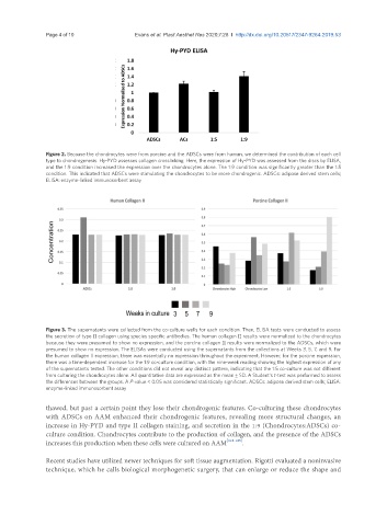
               Figure 2. Because the chondrocytes were from porcine and the ADSCs were from human, we determined the contribution of each cell
               type to chondrogenesis. Hy-PYD assesses collagen crosslinking. Here, the expression of Hy-PYD was assessed from the discs by ELISA,
               and the 1:9 condition increased the expression over the chondrocytes alone. The 1:9 condition was significantly greater than the 1:5
               condition. This indicated that ADSCs were stimulating the chondrocytes to be more chondrogenic. ADSCs: adipose derived stem cells;
               ELISA: enzyme-linked immunosorbent assay


               Figure 3. The supernatants were collected from the co-culture wells for each condition. Then, ELISA tests were conducted to assess
               the secretion of type II collagen using species specific antibodies. The human collagen II results were normalized to the chondrocytes
               because they were presumed to show no expression, and the porcine collagen II results were normalized to the ADSCs, which were
               presumed to show no expression. The ELISAs were conducted using the supernatants from the collections at Weeks 3, 5, 7, and 9. For
               the human collagen II expression, there was essentially no expression throughout the experiment. However, for the porcine expression,
               there was a time-dependent increase for the 1:9 co-culture condition, with the nine-week reading showing the highest expression of any
               of the supernatants tested. The other conditions did not reveal any distinct pattern, indicating that the 1:5 co-culture was not different
               from culturing the chondrocytes alone. All quantitative data are expressed as the mean ± SD. A Student’s t-test was performed to assess
               the differences between the groups. A P-value < 0.05 was considered statistically significant. ADSCs: adipose derived stem cells; ELISA:
               enzyme-linked immunosorbent assay

               thawed, but past a certain point they lose their chondrogenic features. Co-culturing these chondrocytes
               with ADSCs on AAM enhanced their chondrogenic features, revealing more structural changes, an
               increase in Hy-PYD and type II collagen staining, and secretion in the 1:9 (Chondrocytes:ADSCs) co-
               culture condition. Chondrocytes contribute to the production of collagen, and the presence of the ADSCs
               increases this production when these cells were cultured on AAM [118-125] .

               Recent studies have utilized newer techniques for soft tissue augmentation. Rigotti evaluated a noninvasive
               technique, which he calls biological morphogenetic surgery, that can enlarge or reduce the shape and

